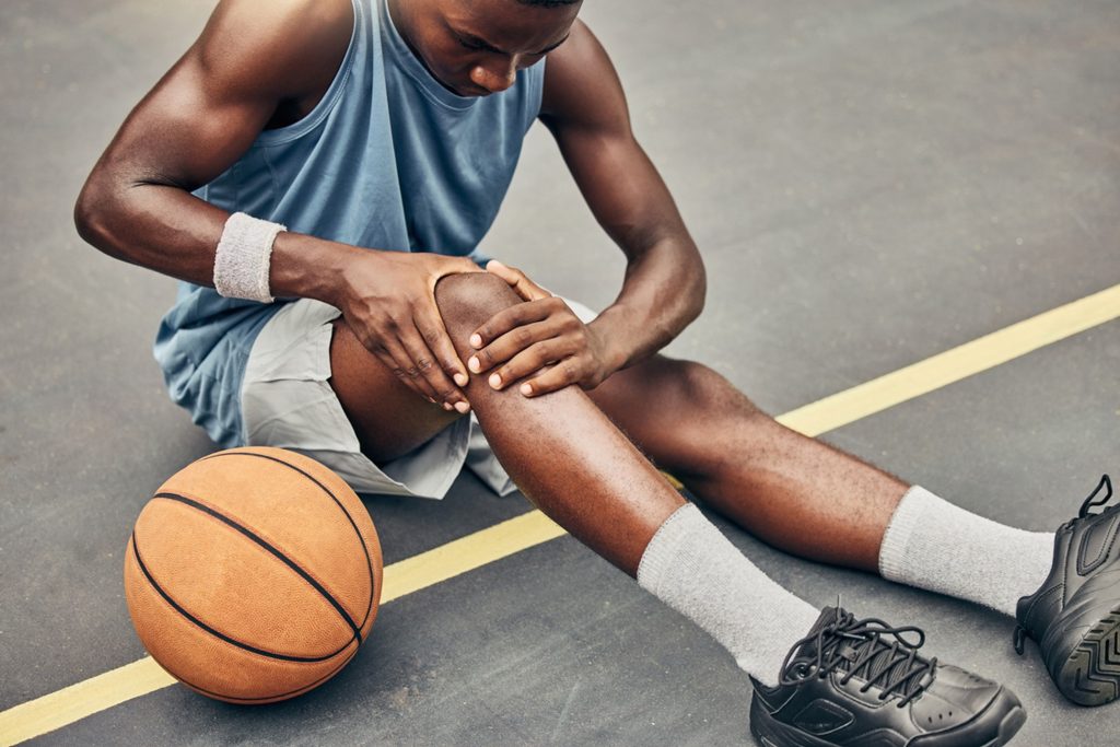
The knee is a complex joint, and the patellar tendon is important for straightening the leg during activities like jumping and running. This tendon (strong fibrous band) connects the kneecap (patella) to the shinbone (tibia). Jumper’s knee happens when this tendon, usually where it attaches to the bottom of the kneecap, gets damaged.
Despite the name “tendinitis,” jumper’s knee is not mainly caused by inflammation. Instead, repeated stress causes tiny tears and wear in the tendon fibres over time. In more serious cases, the tendon can become thicker and more damaged, sometimes with small calcium deposits. Sometimes, nearby tendons around the kneecap can also be affected.
Diagnosis and Risk Factors
Diagnosis
Diagnosis of jumper’s knee starts with a thorough patient history and physical examination. The hallmark symptom is pain just below the kneecap, which worsens with activity—especially jumping, running, or climbing stairs. On examination, there is tenderness at the lower pole of the patella, and pain may be triggered when the patient tries to straighten the knee against resistance.
Imaging studies can help in confirming the diagnosis and rule out other causes of the knee pain:
- X-rays are useful to exclude bone abnormalities, such as bone spurs or fractures.
- Ultrasound can detect tendon swelling, thickening, or partial tears, and is particularly useful for monitoring changes over time.
- MRI (Magnetic Resonance Imaging) provides comprehensive images of the tendon, revealing degenerative changes, partial ruptures, or calcifications. MRI is especially helpful in staging the severity of the condition and planning treatment.
It’s important to note that imaging findings do not always correlate directly with the severity of symptoms; some people may have significant tendon changes on scans but only mild pain.
Risk Factors
Jumper’s knee is primarily caused by repetitive mechanical overload of the patellar tendon. Several risk factors include:
- Overuse and Repetitive Stress: Sports like basketball, volleyball, and football, which involve frequent jumping and sudden changes in direction, place high strain on the patellar tendon.
- Muscle Imbalance: Weakness or tightness in the thigh muscles (quadriceps and hamstrings) can alter knee mechanics and increase tendon load.
- Improper Technique: Poor jumping or landing technique can exacerbate stress on the tendon.
- Inadequate Warm-up: Skipping warm-up exercises decreases tendon flexibility and resilience, making injury more likely.
- Anatomical Factors: Variations such as abnormal patellar height, an increased Q-angle (the angle at which the femur meets the tibia), ligament laxity, or tight leg muscles can contribute to tendon overload.
- Age and Activity Level: While most common in young athletes, jumper’s knee can also affect older adults who remain active, especially if they have reduced flexibility or muscle imbalance.
- Other Factors: Body weight, leg length differences, foot arch height, and training on hard surfaces can also play a role.
Symptoms and Treatment
Jumper’s knee, or patellar tendinopathy, is a condition that causes pain and discomfort around the knee, especially in people who frequently jump, run, or perform intense leg activities.
Symptoms of Jumper’s Knee

The hallmark symptom of jumper’s knee is a dull, aching pain just below the kneecap, where the tendon connects to the bone. Initially, the pain may occur only during physical activity and subside with rest. However, without proper management, it can gradually worsen and persist even during everyday tasks like walking, climbing stairs, or standing for extended periods.
Additional symptoms may include:
- Swelling or tenderness around the lower portion of the kneecap
- Stiffness after periods of inactivity or sleep
- Weakness or instability in the knee when moving
- A burning sensation near the tendon, especially after exercise
If these symptoms linger or worsen, it’s essential to seek medical attention. Delaying care may lead to more severe tendon damage, making recovery more difficult.
Non-Surgical Treatment Options
The main goals of non-surgical treatments are to alleviate pain, support healing, and restore full knee function. Conservative (non-surgical) approaches are generally effective, especially in mild to moderate cases.
- Activity Modification
Reducing or altering activities that aggravate the knee is a critical first step. This doesn’t mean complete rest, but rather avoiding repetitive jumping or running until symptoms subside.
- Cold Therapy and Medication
Applying ice packs several times daily for several days can minimise inflammation and ease discomfort. Nonsteroidal anti-inflammatory drugs (NSAIDs) like ibuprofen may also be used to relieve pain and swelling.
- Physiotherapy
Targeted rehabilitation is essential for tendon recovery. A physiotherapist may recommend:
- Stretching and strengthening exercises
- Eccentric training, which helps rebuild tendon strength
- Techniques to improve jumping and landing form
- Supportive Gear
Knee braces or patellar straps can help reduce strain on the tendon, offering both pain relief and stability during movement.
- Advanced Non-Invasive Therapies
For stubborn cases that don’t respond to standard care:
- Platelet-Rich Plasma (PRP) injections may promote healing by using growth factors from your blood
- Shockwave therapy uses sound waves to stimulate tissue repair
- Corticosteroid injections may be offered sparingly to reduce severe inflammation, though they carry a risk of tendon weakening
Surgical Treatment
Surgery is generally considered when symptoms persist for six months or longer despite extensive conservative treatment. Surgical procedures aim to remove damaged tissue and stimulate tendon healing.
There are two main types:
- Open surgery: The tendon is accessed through a small incision, and degenerated tissue is removed
- Arthroscopic surgery: A minimally invasive approach using a camera and instruments inserted through tiny incisions
Post-surgery, patients typically undergo a structured rehabilitation program to restore strength, flexibility, and function. Full recovery may take several months, but many individuals return to their previous activity level with proper rehab and conditioning.
Prompt identification and management of jumper’s knee are essential to reduce the risk of long-term issues. As previously discussed, non-surgical approaches are often successful in managing early to moderate cases, whereas surgery may be appropriate for more advanced or unresponsive conditions. A well-rounded treatment plan including physiotherapy and patient education can support full recovery, improve functional outcomes, and help individuals safely resume their usual physical activities.
Conclusion
Jumper’s knee is an overuse injury with distinct diagnostic, anatomical, and risk factor profiles. It predominantly affects the patellar tendon at its insertion, is diagnosed clinically and with imaging, and is most common in athletes involved in jumping sports, especially those with high jumping ability and specific landing biomechanics. Prevention and early screening are crucial due to the persistent impact on knee function and athletic careers.
For enquiries and online appointments, send a message to www.DrAyyappanVNair.com/contact
For informative videos related to Shoulder problems and their treatment options, Sports Injuries and other orthopedic conditions, visit our YouTube channel Bangalore Shoulder Institute – https://www.youtube.com/@BangaloreShoulderInstitute

