
Written by: Editorial Team, Bangalore Shoulder Institute
Medically Reviewed by: Dr. Ayyappan V. Nair
Senior Consultant – Shoulder Surgery, Arthroscopy and Sports Trauma
Manipal Hospitals, Bangalore
Last Reviewed: 15th November 2026
Introduction
The menisci are crescent-shaped fibrocartilaginous structures within the knee that play a critical role in load transmission, shock absorption, joint congruency, and knee stability. The medial meniscus is characteristically C-shaped and closely attached to the joint capsule, whereas the lateral meniscus is more circular and mobile because of its relatively fewer capsular attachments.
The discoid meniscus is a congenital morphological variant in which the meniscus is abnormally thickened and disc-shaped rather than having the normal crescent-shaped configuration. It most commonly affects the lateral meniscus and may remain asymptomatic throughout life. However, its abnormal morphology, altered collagen architecture, and potential peripheral instability predispose it to tearing, mechanical symptoms, and functional impairment.
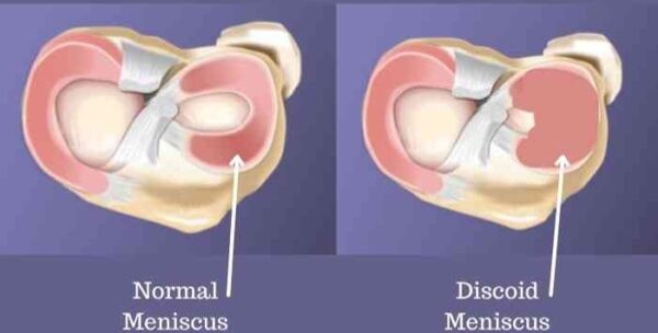
The primary objective of treatment is not simply to remove the abnormal meniscal tissue, but to restore a functional meniscus while preserving as much native tissue as possible.
Epidemiology and pathophysiology
The true incidence of discoid meniscus remains uncertain because many cases are discovered incidentally during imaging or arthroscopy in asymptomatic individuals. The lateral discoid meniscus (DLM) is considerably more common than the medial discoid meniscus (DMM), with a higher reported prevalence in Asian populations.
reported prevalence of approximately 10–13% for DLM in Asian populations, while DMM is extremely rare, with an estimated incidence of 0.1–0.3%. Bilateral involvement is also common in Asian populations, although bilateral anatomical involvement does not necessarily mean that both knees will become symptomatic.
Discoid meniscus develops during early gestational development. Its abnormal morphology is associated with altered collagen formation and an irregular distribution of collagen fibres. These changes may lead to mucoid degeneration and reduced structural integrity, making the meniscus more vulnerable to repetitive microtrauma and tearing.
Histopathological characteristics
Compared with a normal meniscus, the discoid meniscus demonstrates several structural abnormalities:
- Increased meniscal thickness and altered morphology.
- Decreased and disorganised collagen fibres.
- Mucoid degeneration within the meniscal substance.
- Relatively poor vascularity in the inner portion, limiting its healing potential.
In a normal meniscus, collagen fibres are predominantly arranged circumferentially, with radial fibres contributing to structural integrity. This organised architecture allows the meniscus to convert axial compression into circumferential hoop stresses. Disruption of this arrangement may compromise load distribution and contribute to degeneration and tearing in the discoid meniscus.
These histological features are particularly relevant when planning meniscal saucerisation or repair, as preservation of viable tissue and restoration of peripheral stability are essential for long-term function.
Clinical presentation
Discoid meniscus is frequently asymptomatic and may be identified incidentally. Symptoms usually develop when the abnormal meniscus becomes torn, unstable, or mechanically impinged during knee movement.
Common clinical manifestations include:
- Lateral knee pain: the most characteristic pain location in symptomatic DLM.
- Joint effusion: may occur following repetitive activity or a meniscal tear.
- Mechanical symptoms: clicking, locking, catching, or a sensation of giving way.
- Snapping knee syndrome: a palpable or audible clunk, particularly near terminal flexion, classically associated with an unstable Wrisberg variant.
Clinical examination should include lateral joint-line tenderness and appropriate meniscal provocative tests, such as McMurray’s, Apley’s compression, and Thessaly’s tests.
Classification of discoid meniscus
The Watanabe classification remains a widely recognised anatomical classification of discoid lateral meniscus.
Type I — Complete
The meniscus completely covers the lateral tibial plateau. Its broad, disc-like configuration may predispose it to mechanical symptoms and tearing.
Type II — Incomplete
The meniscus is broader than normal but does not completely cover the lateral tibial plateau, retaining a more semilunar configuration.
Type III — Wrisberg variant
The meniscus may have a relatively normal shape but lacks normal posterior meniscal attachments, relying primarily on the meniscofemoral ligament of Wrisberg for posterior connection. This can result in hypermobility and snapping.
This classification is clinically important because morphology alone does not fully define the problem. In particular, the Wrisberg variant requires careful assessment of meniscal stability, as an unstable meniscus may remain symptomatic despite having a relatively normal shape.
Radiological evaluation
Plain radiographs
Radiographs do not directly establish the diagnosis, but certain findings may raise suspicion of a discoid meniscus:
- Widening of the lateral tibiofemoral joint space, approximately 11 mm.
- Squaring of the lateral femoral condyle.
- Cupping of the lateral tibial plateau.
- Hypoplasia of the lateral tibial spine.
These findings should be interpreted in conjunction with the patient’s symptoms and advanced imaging.
Magnetic resonance imaging
MRI is the preferred imaging modality for evaluating discoid meniscus. It helps establish the abnormal morphology, identify associated tears, and assess the peripheral attachments.
Important MRI features include:
- Bow-tie sign
A normally shaped meniscus appears as a bow-tie configuration on a limited number of consecutive sagittal images. In discoid meniscus, the increased anteroposterior extent results in meniscal continuity across more consecutive sagittal sections.
meniscal continuity across more than three consecutive sagittal images with 5-mm slice thickness is a diagnostic criterion.
- Coronal-plane measurements
two useful measurements:
- Maximum meniscal width greater than 15 mm, measured between the peripheral margin and free edge.
- Ratio of minimum meniscal width to maximum tibial width greater than 20%.
- Assessment of associated pathology
MRI should also be reviewed for horizontal cleavage tears, peripheral rim detachment, intrasubstance degeneration, and other associated intra-articular abnormalities. The integrity of the posterior attachments is especially important when instability is suspected.
Management: a meniscus-preserving approach
The management strategy depends on symptoms, tear pattern, meniscal stability, and functional impairment. The presence of a discoid meniscus on MRI is not, by itself, an indication for surgery.
- Conservative treatment
Incidentally identified, asymptomatic discoid menisci can generally be managed with observation and clinical follow-up. Patients should be counselled to seek reassessment if pain, recurrent effusions, locking, or other mechanical symptoms develop.
For symptomatic patients without an urgent mechanical problem, initial management may include activity modification, appropriate analgesia, and rehabilitation directed at restoring strength and function.
Persistent symptoms warrant reassessment for an unstable meniscus or associated tear. The supplied presentation also highlights prolonged symptom duration before partial meniscectomy as a potential risk factor for osteoarthritis, reinforcing the importance of appropriate clinical evaluation and timely management.
- Indications for surgery
Surgical treatment may be considered when there are:
- Persistent symptoms despite appropriate conservative management.
- Severe pain or function-limiting mechanical symptoms.
- A locked knee.
- A symptomatic unstable discoid meniscus or associated tear requiring intervention.
The overarching surgical objective is to create a stable, functional meniscus with a more anatomical contour while preserving viable tissue.
- Arthroscopic saucerisation
Arthroscopic saucerisation, also called reshaping or partial meniscectomy, is the principal tissue-preserving procedure for symptomatic discoid meniscus.
It involves removal of the central excess meniscal tissue to recreate a more normal crescent-shaped configuration. The peripheral rim is preserved to maintain circumferential hoop-stress transmission and meniscal stability.
The key surgical principle is to preserve as much functional meniscal tissue as possible. Excessive resection can compromise load distribution and may increase the risk of later degenerative changes. The amount of rim to preserve should be individualised according to the meniscal dimensions, tissue quality, and stability rather than dictated by a single measurement.
Saucerisation alone is appropriate when the remaining meniscus is stable. If peripheral instability or a significant tear is present, additional repair or stabilisation may be necessary.
Management of associated tears and peripheral instability
Discoid menisci may develop peripheral rim detachment or intrasubstance tears.
Treatment must address both the abnormal morphology and any associated instability.
Peripheral rim detachment
When the peripheral rim is detached but remains repairable, meniscal repair can be performed using an appropriate arthroscopic technique:
- Inside-out repair.
- Outside-in repair.
- All-inside repair.
The choice depends on tear location, tissue quality, accessibility, and the surgeon’s preferred technique. Saucerisation may be combined with repair when the central discoid portion requires reshaping.
Wrisberg variant and hypermobility
A symptomatic Wrisberg variant presents a different surgical challenge. The primary problem may be abnormal posterior attachment and excessive meniscal mobility rather than excessive meniscal width.
In these cases, stabilisation or fixation of the meniscus may be required. Simply reshaping the meniscus without correcting the underlying instability may fail to resolve mechanical symptoms.
Meniscal fixation and centralisation
Meniscopexy involves securing the anterior horn or displaced meniscal tissue to restore a more appropriate position.
It aim to restore meniscal position, improve stability, and preserve the remaining functional tissue. Their selection should be based on the specific pattern of displacement and the integrity of the peripheral attachments.
Postoperative rehabilitation
Rehabilitation should be tailored to the procedure performed and the quality of the meniscal tissue.
Rehabilitation after discoid meniscus surgery
After isolated saucerisation
Immediate weight-bearing and full range of motion as tolerated, followed by icing and quadriceps-strengthening exercises at approximately two weeks.
After saucerisation with meniscal repair
Begin with protected weight-bearing and range-of-motion restrictions. Progression to full weight-bearing and unrestricted range of motion is generally considered around 6–8 weeks in the supplied protocol, depending on healing and the repair performed.
Biomechanical considerations and long-term outcomes
The meniscus is essential for distributing loads across the tibiofemoral joint. Discoid morphology and tearing can alter contact mechanics, while excessive resection may reduce the functional area available for load transmission.
The long-term objective of treatment is to alleviate symptoms without sacrificing the meniscus’s protective function. Persistent symptoms, recurrent tearing, residual instability, and degenerative changes remain important considerations during follow-up.
Key clinical takeaways
An asymptomatic discoid meniscus is generally observed; morphology alone is not an indication for surgery.
MRI helps define the morphology, associated tears, and posterior attachment abnormalities.
Arthroscopic saucerisation aims to restore a more anatomical contour while preserving the functional peripheral rim.
Peripheral detachment and Wrisberg-related hypermobility require specific assessment and may need repair or fixation in addition to saucerisation.
Rehabilitation must distinguish isolated saucerisation from procedures that include meniscal repair.
Conclusion
Discoid meniscus is an important anatomical variant that ranges from an incidental finding to a source of pain, snapping, locking, and meniscal instability. Successful management depends on identifying the cause of symptoms rather than treating the morphology alone.
For symptomatic patients requiring surgery, arthroscopic saucerisation with preservation of the peripheral rim remains a central treatment principle. Associated tears and peripheral instability should be addressed when indicated, particularly in the hypermobile Wrisberg variant. Ultimately, the goal is to achieve a stable, functional meniscus that preserves joint biomechanics and minimises the risk of future degeneration.
For enquiries and online appointments, send a message to www.DrAyyappanVNair.com/contact
For informative videos related to Shoulder problems and their treatment options, Sports Injuries and other orthopedic conditions, visit our YouTube channel Bangalore Shoulder Institute – https://www.youtube.com/@BangaloreShoulderInstitute