Written by: Editorial Team of Dr Ayyappan V Nair
Medically Reviewed by: Dr. Ayyappan V. Nair
Senior Consultant – Shoulder Surgery, Arthroscopy and Sports Trauma
Manipal Hospitals, Bangalore
Last Reviewed: 24th September 2026
Introduction
Articular cartilage injury of the glenohumeral joint is an increasingly recognized component of shoulder pathology, particularly in young, active patients, overhead athletes, and patients with recurrent instability.
Although cartilage lesions may remain clinically silent for a considerable period, their presence can have important long-term implications. Persistent instability, repetitive shear forces and focal cartilage defects may contribute to progressive joint degeneration and ultimately early glenohumeral osteoarthritis, says Dr Ayyappan V Nair, specializing in advanced minimally invasive shoulder procedures and sports injuries.
High-grade articular cartilage injuries have been reported in approximately 17% of high-level overhead throwers [1], while other studies have identified cartilage lesions involving approximately 29% of humeral heads and 15% of glenoids [2].
The challenge is therefore to recognize these lesions early, determine their clinical relevance, address the underlying instability and, where appropriate, preserve or restore the articular surface.
Why Cartilage Matters in the Shoulder
The glenohumeral joint has a relatively thin layer of articular cartilage. The humeral head cartilage has been reported to have an average thickness of approximately 0.89–1.44 mm [3].
Unlike many soft-tissue injuries, articular cartilage has limited intrinsic healing potential. Cartilage defects do not spontaneously heal, and untreated lesions may progressively contribute to degenerative osteoarthritis.
This becomes particularly important in younger patients, in whom the objective is not simply to relieve symptoms but to preserve the native joint for as long as possible.
Focal cartilage defects are particularly relevant in the 25–38-year age group, whereas generalized or diffuse osteoarthritis is more commonly encountered in patients aged 60–70 years and older.
Cartilage Injury and Shoulder Instability
Shoulder instability and cartilage injury frequently coexist.
A recurrently unstable shoulder is repeatedly subjected to:
- Translation and shear
- Impaction
- Rotational loading
- Repetitive microtrauma
- Abnormal joint contact mechanics
Over time, these mechanisms can produce focal chondral injury and contribute to early degenerative changes.
Mechanisms of humeral cartilage injury
The major mechanisms described include:
- Direct axial impact and impaction
- Glenohumeral dislocation producing rotational shear and impaction
- Repetitive microtrauma and torsional shear
- Post-traumatic subchondral vascular insult
Mechanisms of glenoid cartilage injury
Glenoid lesions may result from:
- Glenohumeral dislocation and shear impact
- Repetitive microtrauma during overhead activity
- Eccentric loading and joint incongruity
- Post-traumatic loose-body abrasion
This explains why cartilage assessment should form an integral part of the evaluation of patients with recurrent instability rather than being regarded as an incidental MRI finding.
The GLAD Lesion
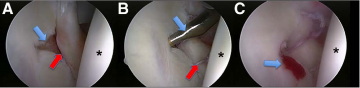
One of the classic cartilage injuries associated with anterior instability is the GLAD lesion—Glenolabral Articular Disruption.
It consists of:
Anterior-inferior labral tear + adjacent glenoid articular cartilage injury
Typical mechanism- forced adduction injury from a position of abduction and external rotation.
Recognition of the cartilage component is important because treating the labral pathology alone may not adequately address the patient’s symptoms when a significant chondral defect is present.
Where Do Cartilage Defects Occur?
The distribution of cartilage lesions is not random.
Humeral head
- Posterolateral region: approximately 60–70%
- Anterosuperior region: approximately 10–15%
- Central/superior region: approximately 15–20%
Glenoid
- Anteroinferior region: approximately 55–65%
- Posterosuperior region: approximately 15–20%
- Central fossa: approximately 15–20%
Understanding these patterns can help the surgeon systematically evaluate the joint during diagnostic arthroscopy.
Clinical Presentation: Why Diagnosis Is Difficult
The symptoms often overlap with other common shoulder disorders, including rotator cuff disease, impingement and instability. Coexisting pathology can further obscure the clinical contribution of the cartilage lesion.
A focal chondral lesion should be considered particularly in patients with:
- Previous shoulder trauma
- Previous shoulder surgery
- Recurrent dislocation or subluxation
- Clicking or catching
- Persistent shoulder pain
- Night discomfort
- Weakness
- Loss of range of motion
The compression-rotation test- a useful clinical manoeuvre
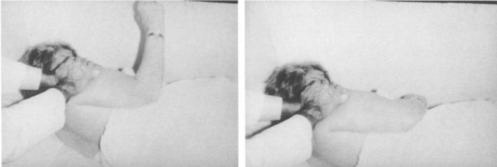
The patient actively internally and externally rotates the shoulder while axial compression is applied across the glenohumeral joint. Pain occurring during the mid-range of motion may suggest a cartilage lesion.
A subacromial injection that eliminates pain attributable to subacromial pathology may potentially increase the specificity of this assessment.
Imaging: MRI Is Useful—But Arthroscopy Remains Crucial
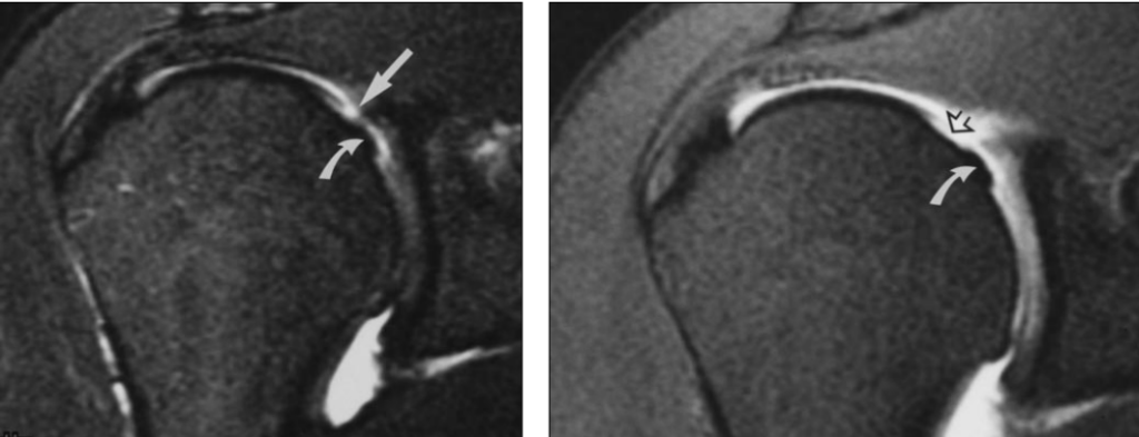
MRI and MR arthrography are valuable because they allow simultaneous assessment of cartilage and associated soft-tissue pathology. However, glenohumeral cartilage lesions can be difficult to accurately characterize on MRI because shoulder cartilage is substantially thinner than cartilage in joints such as the knee.
Arthroscopy
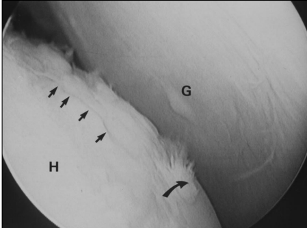
Arthroscopy remains the gold standard for direct assessment.
It allows:
- Direct visualization
- Identification of unstable cartilage
- Assessment of lesion depth
- Measurement of lesion size
- Assessment of surrounding cartilage
- Evaluation of associated labral and instability pathology
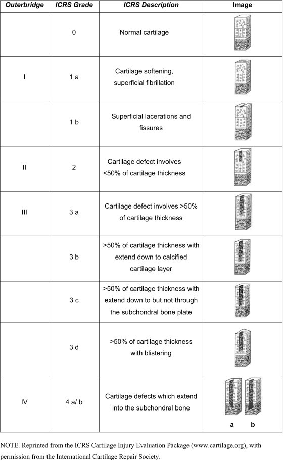
The Outerbridge classification and ICRS classification can be used to characterize cartilage damage.
Treatment: From Palliation to Restoration
Treatment strategies can broadly be divided into:
Palliative → Reparative → Restorative → Reconstructive
Nonoperative Management
Initial management may include:
- NSAIDs
- Physiotherapy
- Activity modification
- Intra-articular viscosupplementation in selected patients
randomized controlled trial by Blaine et al. [4] involving 660 patients in which intra-articular sodium hyaluronate was associated with greater pain relief than saline controls.
High-molecular-weight hyaluronic acid has also been evaluated in patients with shoulder osteoarthritis [5], including a cohort of 78 patients with grade II–IV disease and noted significant improvement in the Constant score (18.2 ± 5.4 and 19.2 ± 5.9, respectively).
Nonoperative treatment, however, does not restore a full-thickness cartilage defect. It is primarily directed toward symptom control and optimization of shoulder function.
Why Not Arthroplasty in the Young Patient?
Shoulder arthroplasty provides excellent symptomatic relief in appropriately selected patients. However, in younger and more active individuals, concerns regarding implant longevity, glenoid component loosening and long-term revision requirements have encouraged the development of joint-preserving procedures.
The goal is therefore to preserve the native joint whenever a biologically and mechanically reasonable solution exists.
1. Palliative Treatment: Debridement
Arthroscopic debridement can be useful for selected small defects, particularly in older patients or when an unexpected cartilage lesion is encountered during surgery.
The objectives are to:
- Remove unstable cartilage
- Eliminate mechanical symptoms
- Reduce catching
- Stabilize the cartilage margins
- Prevent further delamination
However, debridement does not regenerate the lost cartilage and therefore has limited durability for larger full-thickness lesions.
2. Reparative Treatment: Microfracture
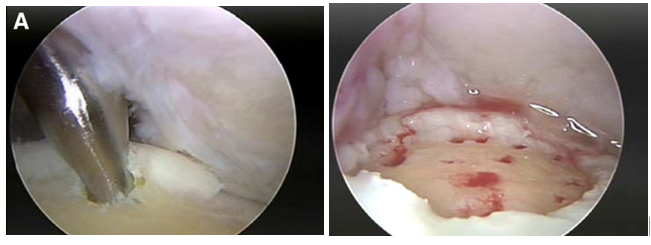
Microfracture is one of the most widely used cartilage reparative procedures.
Multiple small perforations are created in the subchondral bone, typically approximately 3–4 mm apart and 2–3 mm deep, allowing marrow elements to enter the defect.
The resulting marrow clot contains cells and growth factors that contribute to formation of repair tissue, predominantly fibrocartilage.
Clinical outcomes
Millett et al. [6] reported 25 shoulders treated with microfracture, with a mean follow-up of 47 months. Significant reductions in pain and improvements in shoulder function, work, activities of daily living and sports activity were reported.
The major limitation is biological:
Fibrocartilage is not equivalent to native hyaline cartilage.
It is mechanically less durable and may deteriorate over time, particularly in young, high-demand patients.
approximately 70–80% of patients experience significant initial pain and functional improvement, but symptom recurrence may occur after 3–5 years, particularly in athletic individuals.
3. Restorative Procedures
Restorative strategies attempt to replace the defect with tissue that more closely resembles native hyaline cartilage.
These include:
- Osteochondral autograft transfer (OAT/OATS)
- Autologous chondrocyte implantation (ACI)
- Matrix-induced autologous chondrocyte implantation (MACI)
- Bone marrow-derived cellular approaches
Autologous Chondrocyte Implantation
ACI is a two-stage procedure.
Healthy articular cartilage is harvested, chondrocytes are cultured and expanded, and the cells are subsequently implanted into the cartilage defect after several weeks.
In MACI, cultured chondrocytes are seeded onto a collagen membrane, which is then secured directly over the cartilage defect. This approach aims to improve cell retention and reduce leakage compared with traditional techniques.
Studies on MACI demonstrates approximately 75% hyaline-like cartilage regeneration at six months, with good-to-excellent clinical outcomes [7].
Osteochondral Autograft Transfer
OAT is generally considered for smaller focal lesions.
Cylindrical osteochondral grafts containing both cartilage and underlying bone are harvested from relatively non-weight-bearing areas and transferred into the cartilage defect.
The principal advantage is immediate transplantation of mature hyaline cartilage together with its subchondral bone.
MRI evidence [8] of congruent cartilage surfaces and good graft viability and consolidation in most patients in one series, although donor-site pain and recurrent effusion occurred in one patient and required subsequent arthroscopies.
Osteochondral Allograft
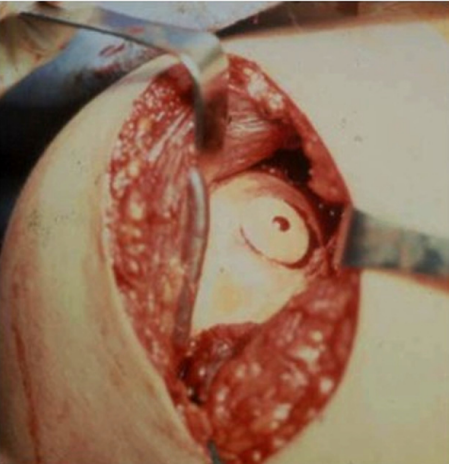
Osteochondral allografts provide an alternative when larger defects make autograft harvesting impractical.
Their principal advantage is that graft size is not restricted by the availability of the patient’s own donor tissue, and there is no donor-site morbidity.
Potential disadvantages include:
- Reduced chondrocyte viability
- Immunogenicity
- Potential disease transmission
Partial Joint Resurfacing
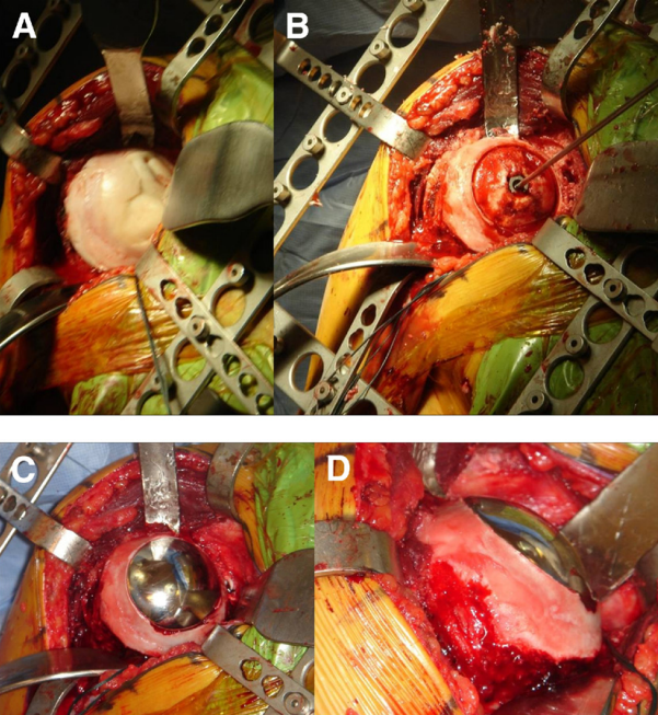
When joint preservation is no longer possible but complete arthroplasty is undesirable, partial resurfacing represents an intermediate option.
Rather than replacing the entire articular surface, a focal portion of the joint is resurfaced.
Stemless prosthetic designs can preserve much of the native proximal humeral anatomy and potentially maintain future reconstructive options.
Biologic Augmentation: BMAC
The expanding field of orthobiologics has introduced bone marrow aspirate concentrate (BMAC) as another potential treatment strategy.
Bone marrow contains mesenchymal stem/stromal cells capable of chondrogenic differentiation and secretion of growth factors. However, the concentration of MSCs in unprocessed bone marrow is extremely low, approximately 0.001–0.02% of nucleated cells.
Processing through density-gradient centrifugation can increase cellular concentration upto 5 times, producing BMAC.
BMAC may be combined with a scaffold to provide a more stable three-dimensional environment for cellular interaction and tissue development.
BMAC Versus Other Cellular Strategies
A 2015 study [9] with 37 patients comparing BMAC vs ACI with patellofemoral chondral lesions was followed up for minimum 3 years. Both groups showed no significant difference in improvement between 2 groups, except for IKDC subjective score (P = 0.015), favored BMAC group. MRI showed complete filling of defects in 76% in MACI and 81% in BMAC
A 2020 meta-analysis [10] involving nine randomized controlled trials and 377 patients found improvements following both bone marrow-derived and adipose-derived MSC treatments, with adipose-derived MSCs showing more consistent improvements in some patient-reported outcomes (VAS and WOMAC scores).
A subsequent 2023 meta-analysis [11] involving 21 studies and 936 patients reported significant improvements (WOMAC, Lysholm and KOOS) at one year with adipose-derived MSCs compared with controls, whereas the results for bone marrow-derived MSCs were less consistent.
Importantly, much of this evidence comes from cartilage literature outside the shoulder, and extrapolation to glenohumeral cartilage defects should therefore be undertaken cautiously.
PRP as an Adjunct
Platelet-rich plasma has also been investigated as an adjunct to cartilage procedures.
systematic review and meta-analysis of 11 studies involving 664 patients [12] comparing PRP + microfracture versus microfracture alone.
PRP augmentation was associated with better outcomes in selected measures, including:
- VAS at 3, 12 and 24 months
- Lysholm scores at 3, 6, 12 and 24 months
- IKDC at 24 months
- Tegner score at three months
Again, much of the available evidence is derived from cartilage literature outside the glenohumeral joint.
Cartilage Defects in the Setting of Instability: Treat the Whole Problem
A key principle in managing cartilage defects associated with instability is that the cartilage lesion should not be considered in isolation.
A patient with recurrent instability may have:
- Labral pathology
- Glenoid bone loss
- Hill-Sachs lesion
- Chondral defect
- Capsular insufficiency
- Rotator cuff pathology
Treating only one component may leave the underlying biomechanical problem unresolved.
For example, persistent instability can continue to expose a repaired or regenerated cartilage surface to abnormal shear and impact.
Therefore:
Joint stabilization and cartilage preservation must often be addressed as complementary objectives.
Case Example: Instability, Bone Loss and a Humeral Cartilage Defect
43-year-old man with recurrent anterior instability and more than 10 episodes of dislocation/subluxation following a previous arthroscopic Bankart repair.
His symptoms had progressed to the point that he could perform only light household activities and was unable to swim or play badminton.
Clinical examination demonstrated a positive apprehension test, with preoperative ASES and Rowe scores of 43 and 30, respectively.
Imaging demonstrated:
- Approximately 15% glenoid bone loss
- An off-track Hill-Sachs lesion
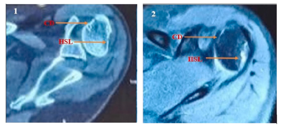
Given the failed Bankart repair and glenoid bone loss, the treatment strategy consisted of:
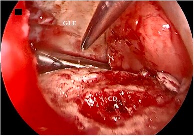
Arthroscopic Latarjet + remplissage + treatment of the superior humeral cartilage defect with BMAC.
Bone marrow was aspirated from the iliac crest, with 60 mL collected and processed using the Arthrex Angel system. The cartilage defect was prepared arthroscopically and the BMAC concentrate was applied to the defect under direct visualization.
Outcome
At one year:
- ASES: 43 → 98
- Rowe: 30 → 95
MRI demonstrated healing of the cartilage defect, complete filling of the Hill-Sachs lesion and satisfactory healing of the capsulotenodesis.
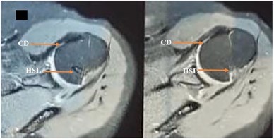
This case illustrates an important contemporary concept:
In a young patient with instability-associated cartilage damage, successful treatment may require simultaneous correction of the mechanical instability and biological management of the cartilage defect.
What Does the Current Evidence Tell Us?
The evidence summarized in the presentation suggests that there is no single universally accepted procedure for every glenohumeral cartilage lesion.
| Treatment | Main role | Principal limitation |
| Debridement | Small/unstable lesions, mechanical symptoms | Does not restore cartilage |
| Microfracture | Small focal full-thickness defects | Fibrocartilage, durability concerns |
| OAT | Small focal lesions | Donor-site limitations |
| OCA | Larger defects | Availability, viability, immunogenicity |
| ACI/MACI | Larger focal defects | Complex, limited shoulder-specific evidence |
| BMAC | Biologic augmentation | Evidence still evolving |
| PRP | Adjunctive biologic treatment | Protocol heterogeneity |
| Partial resurfacing | Focal advanced cartilage loss | Prosthetic procedure |
| Arthroplasty | Advanced degenerative disease | Implant longevity and revision concerns in young patients |
OAT/OCA can demonstrate high integration rates, while microfracture may provide good initial symptom relief but can have durability limitations.
The Young Athlete: Why Early Recognition Matters
Young athletes and high-demand patients represent a particularly important population.
The combination of:
Traumatic injury + instability + repetitive loading + focal cartilage damage
may initiate a cycle of progressive joint damage.
key principles:
- The humeral head has relatively thin articular cartilage.
- Chronic instability may contribute to early glenohumeral osteoarthritis.
- Active patients and athletes have increased risk of traumatic cartilage lesions.
- Cartilage defects may remain clinically occult until more advanced stages.
- Early recognition and joint-preserving treatment should therefore be considered in appropriate young, active patients.
Conclusion
Cartilage defects of the glenohumeral joint are an important but sometimes under-recognized cause of persistent shoulder symptoms, particularly in young active patients and those with recurrent instability.
The key principles are:
- Think about cartilage injury in recurrent instability and post-traumatic shoulders.
- Do not rely exclusively on MRI; diagnostic arthroscopy remains crucial.
- Assess the entire joint—including labrum, bone loss, Hill-Sachs lesion and cartilage.
- Small lesions may be managed with debridement or microfracture.
- Larger focal lesions may require restorative procedures such as OAT, OCA or ACI/MACI.
- Biologic approaches such as BMAC and PRP are promising but require further shoulder-specific evidence.
- In instability-associated cartilage lesions, correcting the mechanical instability is as important as treating the cartilage defect.
- In young patients, joint preservation should remain the overarching goal whenever biologically and mechanically appropriate.
The modern approach to shoulder cartilage disease is therefore not simply “repair the cartilage.”
It is:
Restore stability, optimize joint mechanics, preserve native cartilage whenever possible, and use biological restoration strategically in appropriately selected patients.
For informative videos related to Shoulder problems and their treatment options, Sports Injuries and other orthopedic conditions, visit our YouTube channel Bangalore Shoulder Institute – https://www.youtube.com/@BangaloreShoulderInstitute
REFERENCES
- Paley, K.J., Jobe, F.W., Pink, M.M., Kvitne, R.S. and ElAttrache, N.S. (2000), Arthroscopic Findings in the Overhand Throwing Athlete: Evidence for Posterior Internal Impingement of the Rotator Cuff. Arthroscopy, 16: 35-40. https://doi.org/10.1016/S0749-8063(00)90125-7
- Guntern DV, Pfirrmann CW, Schmid MR, Zanetti M, Binkert CA, Schneeberger AG, Hodler J. Articular cartilage lesions of the glenohumeral joint: diagnostic effectiveness of MR arthrography and prevalence in patients with subacromial impingement syndrome. Radiology. 2003 Jan;226(1):165-70. doi: 10.1148/radiol.2261012090. PMID: 12511686.
- Depalma AA, Gruson KI. Management of cartilage defects in the shoulder. Curr Rev Musculoskelet Med. 2012 Sep;5(3):254-62. doi: 10.1007/s12178-012-9131-1. PMID: 22628177; PMCID: PMC3535081.
- Blaine T, Moskowitz R, Udell J, Skyhar M, Levin R, Friedlander J, Daley M, Altman R. Treatment of persistent shoulder pain with sodium hyaluronate: a randomized, controlled trial. A multicenter study. J Bone Joint Surg Am. 2008 May;90(5):970-9. doi: 10.2106/JBJS.F.01116. PMID: 18451387.
- Di Giacomo G, de Gasperis N. Hyaluronic Acid Intra-Articular Injections in Patients Affected by Moderate to Severe Glenohumeral Osteoarthritis: A Prospective Randomized Study. Joints. 2017 Aug 11;5(3):138-142. doi: 10.1055/s-0037-1605389. PMID: 29270542; PMCID: PMC5738473.
- Millett PJ, Huffard BH, Horan MP, Hawkins RJ, Steadman JR. Outcomes of full-thickness articular cartilage injuries of the shoulder treated with microfracture. Arthroscopy. 2009 Aug;25(8):856-63. doi: 10.1016/j.arthro.2009.02.009. PMID: 19664505.
- Zheng MH, Willers C, Kirilak L, Yates P, Xu J, Wood D, Shimmin A. Matrix-induced autologous chondrocyte implantation (MACI): biological and histological assessment. Tissue Eng. 2007 Apr;13(4):737-46. doi: 10.1089/ten.2006.0246. PMID: 17371156.
- Scheibel M, Bartl C, Magosch P, Lichtenberg S, Habermeyer P. Osteochondral autologous transplantation for the treatment of full-thickness articular cartilage defects of the shoulder. J Bone Joint Surg Br. 2004 Sep;86(7):991-7. doi: 10.1302/0301-620x.86b7.14941. PMID: 15446525.
- Gobbi A, Chaurasia S, Karnatzikos G, Nakamura N. Matrix-Induced Autologous Chondrocyte Implantation versus Multipotent Stem Cells for the Treatment of Large Patellofemoral Chondral Lesions: A Nonrandomized Prospective Trial. Cartilage. 2015 Apr;6(2):82-97. doi: 10.1177/1947603514563597. PMID: 26069711; PMCID: PMC4462249.
- Han X, Yang B, Zou F, Sun J. Clinical therapeutic efficacy of mesenchymal stem cells derived from adipose or bone marrow for knee osteoarthritis: a meta-analysis of randomized controlled trials. J Comp Eff Res. 2020 Apr;9(5):361-374. doi: 10.2217/cer-2019-0187. Epub 2020 Mar 6. PMID: 32141308.
- Muthu S, Patil SC, Jeyaraman N, Jeyaraman M, Gangadaran P, Rajendran RL, Oh EJ, Khanna M, Chung HY, Ahn BC. Comparative effectiveness of adipose-derived mesenchymal stromal cells in the management of knee osteoarthritis: A meta-analysis. World J Orthop. 2023 Jan 18;14(1):23-41. doi: 10.5312/wjo.v14.i1.23. PMID: 36686284; PMCID: PMC9850793.
- Mahamat Abdramane A, Abdirahman A, Opoku M, Fang M, Li Y, Yang X, Xiao W. Effectiveness of platelet-rich plasma plus microfracture compared with microfracture alone in the treatment of knee cartilage lesions: a systematic review and meta-analysis of comparative studies. Int J Surg. 2026 Jan 12;112(4):10504–22. doi: 10.1097/JS9.0000000000004610. Epub ahead of print. PMID: 41427522; PMCID: PMC13105750.