
Written by: Editorial Team, Bangalore Shoulder Institute
Medically Reviewed by: Dr. Ayyappan V. Nair
Senior Consultant – Shoulder Surgery, Arthroscopy and Sports Trauma
Manipal Hospitals, Bangalore
Last Reviewed: 15th November 2026
The shoulder dislocates forward when the arm is forced up and back, as in a tackle, a fall onto an outstretched hand or a heavy landing in a throwing position. The ball of the humerus slides out in front of a socket that is shallow by design, tearing soft tissue and sometimes chipping bone on the way. What the dislocation damages on its way out determines whether the shoulder settles afterwards or keeps slipping.
How the Joint Gives Way at the Front
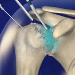
The glenoid, the socket of the shoulder, is a shallow dish that covers only a fraction of the humeral head. A rim of fibrocartilage called the labrum deepens it. The front band of the inferior glenohumeral ligament is anchored to that rim and holds the ball in place when the arm is lifted out to the side and turned outwards. A sudden force in that position overloads the ligament, the labrum peels away from the bone, and the humeral head escapes forward and downward. With the restraint detached, the joint has less to resist the next episode.
What a Dislocation Can Damage
Several structures can be injured in the same event, and each one changes the treatment plan:
- Bankart lesion: The labrum and its attached ligament tear off the front and lower edge of the socket. This is the most common injury and removes the main brake against forward movement.
- Bony Bankart lesion: A fragment of the socket rim breaks away with the labrum. Lost bone shortens the arc of the socket, so the ball has less to push against in later episodes.
- Hill-Sachs lesion: The back of the humeral head is dented as it strikes the socket rim. If the dent lines up with the rim when the arm is raised, the head can catch and lever out again.
- Rotator cuff tear: This is more frequent after the age of 40. Weakness in lifting the arm once the first pain eases is the usual clue.
- Axillary nerve injury: Numbness over the outer upper arm and a weak deltoid point to a stretched nerve. It often recovers over weeks to months but should be documented early.
Numbness or weakness in the hand, or a cold and pale hand, needs emergency care, since the nerves and blood vessels around the shoulder can occasionally be injured.
Traumatic Instability and the Loose-Jointed Shoulder
Traumatic instability follows a definite injury and leaves structural damage that scans can show. Atraumatic or multidirectional instability is a different condition. It is seen in people with naturally lax ligaments, often in both shoulders, where the joint slips with little force and in more than one direction. A torn labrum usually needs surgical repair once dislocations recur, whereas the loose-jointed shoulder responds first to targeted physiotherapy, and operating on it as though it were a torn labrum risks a poor result.
Age and Activity Shape the Risk of a Second Dislocation
Teenagers and young adults who play contact or overhead sport carry the highest risk of dislocating again, and several studies report that a majority redislocate after non-operative treatment. The risk falls steadily with age. After 40 the ligaments tend to hold while the rotator cuff tears, so persistent weakness and stiffness become the main concerns. For parents, a first dislocation in a young athlete deserves a prompt specialist review, because each recurrence can wear more bone from the socket and the humeral head.
What Imaging Needs to Answer
X-rays taken before and after reduction confirm the position of the joint and show fractures. MRI, often with contrast injected into the joint, shows the labrum, ligaments and rotator cuff. CT with three-dimensional reconstruction measures the bone lost from the socket and the size of the Hill-Sachs dent. Those measurements steer the decision between a soft tissue repair and a bone procedure. The examination adds the apprehension test, where moving the arm towards the injury position produces a sense that the shoulder is about to slip, along with a sensation check over the outer upper arm.
Treatment: Matching the Operation to the Damage
After reduction, most people wear a sling for comfort for a short period and then start physiotherapy to strengthen the rotator cuff and shoulder blade muscles. This approach suits older patients, lower-demand activity and shoulders without significant bone loss. Physiotherapy builds dynamic control but cannot reattach a torn labrum, so surgery is considered after recurrent dislocations. Many surgeons also offer early stabilisation to young athletes in contact sport after a first dislocation.
Procedure | Best suited to | Main trade-off |
Arthroscopic Bankart repair | Torn labrum with little bone loss | Keyhole surgery with a higher recurrence risk when bone loss is significant or the sport involves collisions |
Bankart repair with remplissage | Labral tear plus a Hill-Sachs dent that catches on the rim | Tendon and capsule fill the dent, which may slightly reduce outward rotation |
Latarjet procedure | Significant socket bone loss, failed repair, high-demand contact sport | Moves a piece of coracoid bone to the front of the socket and adds a sling effect from the attached tendon, but it is a larger operation with risks including nerve injury and graft or screw problems |
Bone block graft | Bone loss when a Latarjet is unsuitable | Rebuilds the socket with donor bone and is used in selected cases |
Recovery and Return to Sport
After surgery the arm rests in a sling for a few weeks, commonly three to six, while the repair heals. Movement returns first, strengthening follows, and sport-specific drills come last. Return to sport is commonly around six months, and contact or collision sport can take longer. Clearance depends on strength, movement and confidence in the shoulder as well as time, because returning early exposes a healing repair to the same force that damaged it originally.
A shoulder that has been put back in place can feel nearly normal while a torn labrum and a dented humeral head remain. Each further dislocation can remove more bone from the socket, and once enough is gone a keyhole repair no longer holds. A scan and a specialist opinion after the first dislocation keep the simpler options available.
For enquiries and online appointments, send a message to www.DrAyyappanVNair.com/contact
For informative videos related to Shoulder problems and their treatment options, Sports Injuries and other orthopedic conditions, visit our YouTube channel Bangalore Shoulder Institute – https://www.youtube.com/@BangaloreShoulderInstitute