
Written by: Editorial Team, Bangalore Shoulder Institute
Medically Reviewed by: Dr. Ayyappan V. Nair
Senior Consultant – Shoulder Surgery, Arthroscopy and Sports Trauma
Manipal Hospitals, Bangalore
Last Reviewed: 15th November 2026
The Anterior Cruciate Ligament (ACL) is one of the most vital structures stabilizing the knee joint.
- Macroanatomy and Geometry [1]
The ACL is a dense band-like structure of connective tissue.
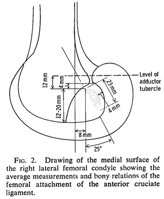
- Femoral Attachment: The femoral attachment forms a vertically disposed semicircle located on the posterior part of the inner surface of the lateral femoral condyle, measuring roughly 11 to 24 mm across.
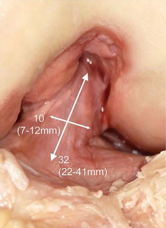
- Course and Dimensions: From its origin on the femur, the ACL runs anteriorly, medially, and distally toward the tibia. Its total length ranges from 22 to 41 mm (with a mean of 32 mm), while its width ranges between 7 and 12 mm.
- Cross-Sectional Shape & Area: The cross-sectional shape is irregular and changes depending on the angle of knee flexion. The cross-sectional area progressively increases from the femur down to the tibia [2]:
- Proximally:
- Mid-proximally:
- Mid-substance:
- Mid-distally:
- Distally:
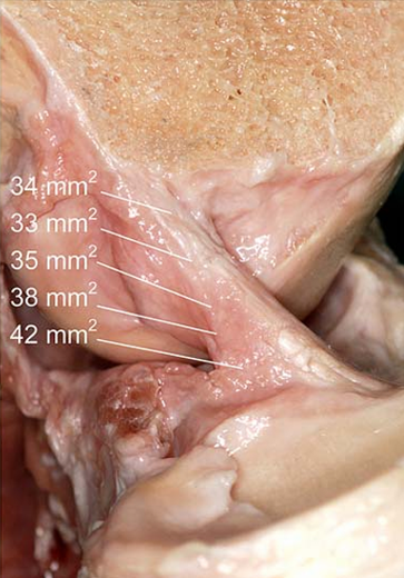
- Tibial Attachment: As the ACL fibers approach the tibia, they fan out and attach to a wide, depressed fossa located anterior and lateral to the medial tibial spine. This tibial footprint is approximately 11 mm wide and 17 mm in the anteroposterior direction.
- Functional Bundles and Kinematics
While some researchers have divided the ACL into three bands, the two-bundle model (pioneered by Girgis et al.) [1] remains the most widely accepted representation of ACL function. These two primary functional components are:
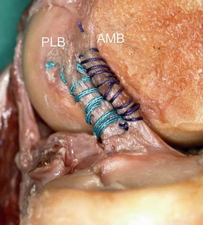
- Anteromedial Bundle (AMB): Originates at the most anterior and proximal aspect of the femoral attachment and inserts at the anteromedial aspect of the tibia.
- Posterolateral Bundle (PLB): Originates at the postero-distal aspect of the femur and inserts at the posterolateral aspect of the tibia.
How They Move: Non-Isometric Behavior [3]
During passive knee flexion, the two bundles experience distinct length patterns rather than moving isometrically:
- Extension: The fascicles run fairly parallel, with a significant length difference between the AMB (34 mm) and PLB (22.5 mm).
- Flexion: As the knee flexes, the ligament undergoes a slight lateral rotation around its longitudinal axis, and the AMB begins to spiral around the rest of the ligament.
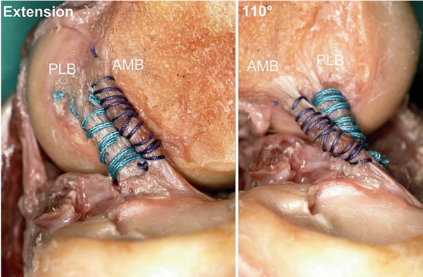
- Length Changes: According to Hollis et al., the AMB lengthens and tightens during flexion (increasing by ~5% at 30 degrees and ~12% at 90 degrees). Conversely, the PLB shortens and becomes slack, decreasing in length by 14% at 30 degrees and 32% at 90 degrees of flexion.
- Innervation, Proprioception, and Blood Supply
- Vascularization: The primary blood supply comes from the middle genicular artery (MGA), including a large posteriorly descending branch directed to the upper tibia. Notably, the proximal part of the ACL is significantly better vascularized than its distal portion.
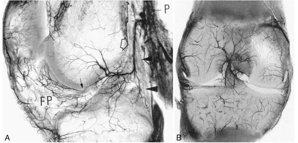
- Innervation & The “ACL Reflex”: Nerve fibers from the posterior articular branches of the tibial nerve penetrate the capsule and accompany blood vessels into the ligament. Mechanoreceptors (Ruffini, Pacini, and Golgi-like receptors) provide vital proprioceptive feedback and provide the afferent arc for signaling knee postural changes. Deformations within the ACL influence muscle spindle output through the fusimotor system, directly affecting motor activity in muscles surrounding the knee—a phenomenon known as the ACL reflex.
- Clinical Examination and Diagnostic Accuracy
Accurately diagnosing an ACL tear relies heavily on specialized physical examination tests. A blinded, cross-sectional evaluation highlights the sensitivity and specificity of the top clinical tests: [4]
Clinical Test | Sensitivity | Specificity |
Pivot Shift Test | 98% | 24% |
Anterior Drawer Test | 92% | 91% |
Lachman Test | 85% | 94% |
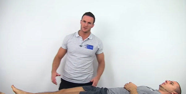
Lachman test
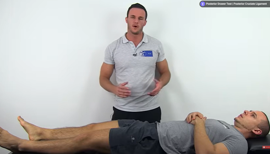
Anterior Drawer Test
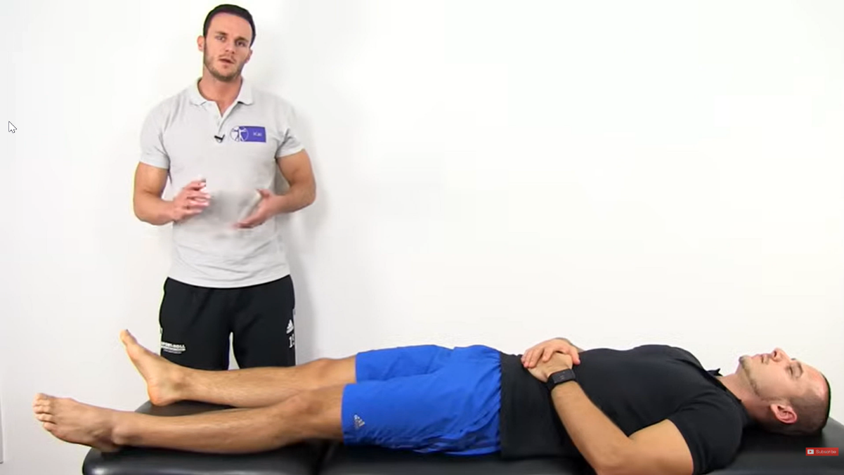
Pivot Shift Test
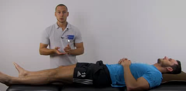
Lever test [5]
For enquiries and online appointments, send a message to www.DrAyyappanVNair.com/contact
For informative videos related to Shoulder problems and their treatment options, Sports Injuries and other orthopedic conditions, visit our YouTube channel Bangalore Shoulder Institute – https://www.youtube.com/@BangaloreShoulderInstitute
REFERENCES
- Girgis FG, Marshall JL, Monajem A. The cruciate ligaments of the knee joint. Anatomical, functional and experimental analysis. Clin Orthop Relat Res. 1975 Jan-Feb;(106):216-31. doi: 10.1097/00003086-197501000-00033. PMID: 1126079.
- Harner CD, Livesay GA, Kashiwaguchi S, Fujie H, Choi NY, Woo SL. Comparative study of the size and shape of human anterior and posterior cruciate ligaments. J Orthop Res. 1995 May;13(3):429-34. doi: 10.1002/jor.1100130317. PMID: 7602404.
- Hollis JM, Takai S, Adams DJ, Horibe S, Woo SL. The effects of knee motion and external loading on the length of the anterior cruciate ligament (ACL): a kinematic study. J Biomech Eng. 1991 May;113(2):208-14. doi: 10.1115/1.2891236. PMID: 1875695.
- Kulwin RL, Schmidt GJ, Snyder DA, Klitzman RG. Clinical Examination in the Diagnosis of Anterior Cruciate Ligament Injury: A Blinded, Cross-sectional Evaluation. J Am Acad Orthop Surg Glob Res Rev. 2023 Feb 8;7(2):e22.00123. doi: 10.5435/JAAOSGlobal-D-22-00123. PMID: 36758231; PMCID: PMC9915950.
- Lelli A, Di Turi RP, Spenciner DB, Dòmini M. The “Lever Sign”: a new clinical test for the diagnosis of anterior cruciate ligament rupture. Knee Surg Sports Traumatol Arthrosc. 2016 Sep;24(9):2794-2797. doi: 10.1007/s00167-014-3490-7. Epub 2014 Dec 25. PMID: 25536951.