

Ayyappan V. Nair, D.Ortho, D.N.B., M.N.A.M.S., Pramod Kumar Mohan, M.S. Ortho.,
Ajit Jangale, M.S., M.R.C.S., Pavan Krishna, M.S. Ortho.,
Prince Shanavas Khan, D.Ortho, M.S. Ortho., Maythilisharan Rambhojun, M.S. Ortho., and Sreejith Thampy, M.S. Ortho.
- Arthroscopy Techniques – 2024
LINK: https://www.sciencedirect.com/science/article/pii/S2212628724002792
This article presents an arthroscopic technique for treating chronic acromioclavicular (AC) joint dislocations by reconstructing both coracoclavicular (CC) and acromioclavicular ligaments using an autogenous gracilis tendon graft reinforced with FiberTape and secured with a dog button. Unlike conventional open procedures, this method minimizes soft-tissue dissection, reduces implant usage, and enhances graft healing by placing it through a coracoid tunnel. The technique employs two clavicular tunnels to reproduce the anatomic double-bundle CC ligaments, while tendon ends are configured in a figure-of-eight for AC ligament reconstruction, restoring both vertical and horizontal stability. Patient positioning is in a semi-beach-chair setup with graft harvested from the ipsilateral knee. The procedure includes diagnostic arthroscopy, graft preparation, coracoid and clavicle tunnel drilling, graft passage, and fixation. Postoperatively, the shoulder is immobilized for 6 weeks with gradual rehabilitation, and patients can return to sports after 6 months. Compared with traditional methods, this approach addresses high complication rates of open reconstructions and risks of graft stretching, tunnel widening, or implant failure. The FiberTape reinforcement and dog button provide additional stability, reducing recurrence. The described technique aims to restore near-native biomechanics with fewer complications, offering a robust and reproducible option for managing chronic AC joint dislocations.
| A |
cromioclavicular (AC) joint injuries are more methods. The management of type III AC joint injuries common among young males, especially athletes remained controversial until type III injuries were
| with high-impact risks such as contact sports, football, ice hockey, and wrestling, with an overall incidence reported to be 9.2 injuries per 1,000 person-years in younger athletes.1,2 It accounts for 12% of all shoulder girdle injuries3 and mostly occurs due to a direct fall on the shoulder with an adducted arm position. Type I and II AC joint injuries are treated conservatively with good to excellent outcomes, whereas type IV to VI AC joint injuries are managed with surgical | subdivided into type III A (horizontally stable) and type B (horizontally unstable). The latter type III B is managed surgically to avoid the chronic instability that leads to pain and dysfunction.4 Most of the injuries can be managed with repair techniques when presented within 3 weeks due to biological healing properties. The injuries lasting more than 3 weeks require anatomic reconstruction of the ligaments using biological grafts.5,6 However, the complication rates of surgically treated AC joint injuries are high.7 Recently, arthroscopic techniques have been |
described that minimize soft tissue dissection and improve the healing rates along with addressing
concomitant intra-articular pathologies.8,9 The complication rates following the arthroscopic approach for ligament reconstruction range from 12.5% to 27.1%.10 Restoring both horizontal and vertical stability provides excellent clinical outcomes, reducing the incidence of postoperative horizontal instability.11 We describe a modification of the arthroscopic coracoclavicular ligament reconstruction using a single coracoid tunnel with
Arthroscopy Techniques, Vol 14, No 1 (January), 2025: 103154 e1
a gracilis autograft reinforced with FiberTape (Arthrex), providing robust reconstruction with less incidence of postoperative graft stretching.
Patient Positioning
The patient is in a beach-chair position with the arm draped separately without a traction device. The ipsilateral knee is draped for tendon graft harvesting (Fig 1). We use intraoperative C-arm fluoroscopy positioned on the opposite side of the patient. Patients are anesthetized with general anesthesia and a regional nerve block under hypotensive anesthesia.
Surgical Technique
Diagnostic Arthroscopy
Using a 4-mm 30 arthroscope (Stryker) and standard posterior soft tissue portal, a diagnostic glenohumeral arthroscopy is done. Any intra-articular pathologies are addressed (Video 1).
Gracilis Tendon Harvest and Preparation
The gracilis tendon graft is harvested from the ipsilateral knee and the prepared tendon is augmented with a FiberTape (Arthrex) internal brace. A dog button (Arthrex) is placed in the middle of the prepared graft over the FiberTape, making the tendon graft doublestranded (Fig 2).
Coracoid Exposure
Rotator interval portal is made with an outside-in technique, viewing from the posterior soft tissue portal. The rotator interval is exposed using a radiofrequency probe (Stryker). The under surface of the coracoid is exposed and skeletonized of the soft tissue attachments (Fig 3). Further coracoid exposure is done through the anterolateral portal. The arthroscope is shifted to the anterolateral portal by railroading over a switching stick.
Pectoralis Minor Tendon Release
The midaxillary portal is made using the inside-out technique and the arthroscope is shifted to the midaxillary portal. The midaxillary portal is used as the visualization portal for further surgery. The pectoralis major tendon is elevated using a switching stick from the anterolateral portal for good visualization of the anterior surface of the coracoid. The pectoralis minor tendon is exposed and released from the medial side of
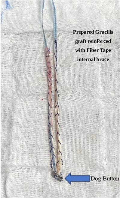
Fig 2. The harvested gracilis tendon is prepared on the back table. The prepared gracilis autograft is reinforced with the FiberTape (Arthrex) and the dog button is incorporated in the center of the graft over the FibreTape. the coracoid, working from the trans-pectoralis major portal, made medial to the conjoint tendon.
Coracoid Drilling and Passage of Suture Loop
Visualizing from the midaxillary portal, the supracoracoid portal is made using the inside-out technique. The drilling sleeve is passed through the coracoid portal and aimed for the knee of the coracoid. The coracoid tunnel is made with calibrated guidewire, and sequential reaming is done for matching the graft diameter, making sure the graft diameter is kept less than 6 mm (Fig 4). The nitinol wire loop is passed through the coracoid tunnel and
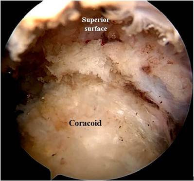
Fig 3. Viewing with the 4-mm 30 arthroscope from the midaxillary portal, with the patient in a semi-beach chair position with the right shoulder showing the upper surface of the coracoid after the release of the soft tissue attachments, working from the anterolateral and trans-pectoralis portals. The coracoid is skeletonized and the pectoralis minor tendon is released taking care of the musculocutaneous nerve.
parked outside the anterolateral portal. The nitinol wire loop is replaced with No. 2 polydioxanone (PDS) suture loop.
Distal Clavicle Exposure and Resection
The acromion and the lateral third of the clavicle are exposed with meticulous dissection. The distal end of the clavicle measuring 10 mm is resected using the bone saw and the superior surface is rasped to prevent soft tissue irritation from sharp edges.
Clavicular Tunnel Preparation and Passage of Suture Loops
Two tunnels are drilled from the posterior to anterior direction over the superior surface of the clavicle.
Two tunnels are made approximately 25 mm and 45 mm from the distal end of the clavicle to re-create the anatomic double-bundle coracoclavicular ligaments (Fig 5). The drill holes are made with cannulated 3.2-mm drill bites over predrilled K-wires. An 18-gauge wide-bore needle is used to shuttle a nonabsorbable PDS suture through the clavicular drill holes. The PDS suture ends are visualized from the midaxillary portal and retrieved outside the anterolateral portal.
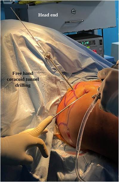
Fig 4. Viewing with the 4-mm 30 arthroscope from the midaxillary portal, with the patient in a semi-beach chair position for the right shoulder, and the drill sleeve is passed from the supracoracoid portal. The free hand coracoid tunnel is drilled with a calibrated guidewire with a cannulated drill sleeve centered over the knee of the coracoid from the supracoracoid portal, with the patient in a semi-beach chair position for the right shoulder. The coracoid tunnel is positioned to be in the center-center position for optimum tunnel positioning.
Passage of Gracilis Tendon Graft and Securing of Graft Ends
The FiberTape ends are shuttled through the nitinol loop and retrieved through the coracoid portal. The color-coded ends are retrieved along with the appropriate PDS suture limb and passed through the clavicle tunnels separately to avoid the soft tissue window between the sutures (Fig 6). Each end of the graft is shuttled through the clavicle tunnels and retrieved through the soft tissue incision made over the lateral end of the clavicle. The ends of the FiberTape are pulled through the clavicular tunnels until the dog button is
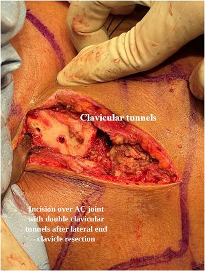
Fig 5. The lateral end of the clavicle along with the acromion is exposed with an incision over the clavicle. Two tunnels, drilled from the posterior to anterior direction over the superior surface of the clavicle, are made approximately 25 mm and 45 mm from the distal end of the clavicle with a 3.2-mm cannulated drill bit, with the patient in a semi-beach-chair position for the right shoulder.
seated well under the coracoid surface (Figs 7-9). The clavicle is reduced with a bone spike placed under the acromion as a fulcrum and under fluoroscopy control, making sure to obtain an anatomic reduction of the joint. The tendon ends are secured over the clavicle surface with a simple overhand throw and fixed with an absorbable 2.0 Vicryl (Ethicon) sutures, cutting them close and making sure to avoid knot irritation (Fig 10).
Acromioclavicular Ligament Reconstruction
The tendon ends are passed through the tunnel in the acromion, which is drilled with a cannulated 3.2mm drill bit in the posterior to anterior direction. The tendon ends are secured together in a figure-of-8 configuration reconstructing the acromioclavicular ligament (Fig 11). The final arthroscopic evaluation demonstrates the dog button seated flush over the coracoid under the surface with the gracilis graft with
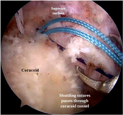
Fig 6. Viewing from the midaxillary portal with a 4-mm 30 arthroscope with the patient in a semi-beach-chair position for the right shoulder, the gracilis graft reinforced with the FiberTape (Arthrex) is passed through the coracoid tunnel, and each end of the FiberTape is shuttled along the polydioxanone suture loop passed through the clavicular tunnel. The graft is passed through the anterolateral portal with visualization from the midaxillary portal.
adequate tension on the graft. The final fluoroscopic evaluation shows a well-reduced AC joint (Figs 12-14).
Postoperative Rehabilitation
The shoulder is immobilized with a shoulder immobilizer for 6 weeks. Active elbow range of motion and hand grips along with shoulder shrugs are initiated soon after the patient is comfortable after the surgery. The passive range of movement of the shoulder is started after 2 weeks to attain full forward flexion by the end of the sixth week. Active strengthening and range-of-motion exercises are taught to the patient after 6 weeks and continued until the patient achieves full power and range of motion of the shoulder. Patients are allowed to be involved in active sports after a period of 6 months, after the patient is evaluated by a shoulder-specific sports physiotherapist.
Discussion
Surgical treatment of chronic AC joint injuries remains a challenging entity due to a paucity in the clear consensus of the treatment protocol. Many techniques have been described in the literature, but superiority of one technique over the other is still not established.
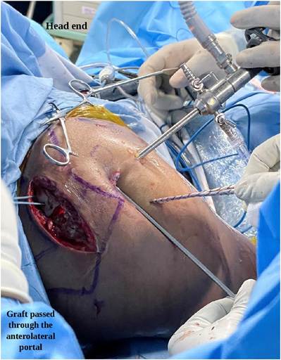
Fig 7. Viewing with the 4-mm 30 arthroscope from the midaxillary portal, with the patient in a semi-beach-chair position for the right shoulder, the gracilis graft reinforced with the FiberTape (Arthrex) with the dog button is passed through the anterolateral portal. The dog button is incorporated in such a way that it is placed in the center of the total graft length.
Complication rates with open techniques are high, including excessive soft dissection around the coracoid, implant failures necessitating future removal, excessive scarring, and loss of reduction. Arthroscopic techniques have advantages of addressing concomitant glenohumeral pathologies associated with AC joint injuries estimated around 30%.12
Chronic AC joint injuries managed with free graft reconstruction have been shown to have better outcomes and a stable construct when compared with the modified Weaver-Dunn procedure. Free graft reconstruction provides both vertical and horizontal stability, ensuring good stability along with superior clinical and radiographic outcomes.10,13 Double-bundle reconstruction of the coracoclavicular ligament provides superior vertical stability compared to single-bundle reconstruction.14
Clavicular and coracoid fractures following AC joint reconstruction procedures have been described in the literature. Techniques involving looping of graft around
Fig 8. Viewing with the 4-mm 30 arthroscope from the midaxillary portal, with the patient in a semi-beach-chair position for the right shoulder, the 2 limbs of the graft ends are reinforced with the FiberTape (Arthrex) shuttled through the clavicular tunnels from the anterolateral portal. The color coding of the passing sutures helps with the appropriate passage of the graft ends through the clavicular tunnels.
Fig 10. The surgical picture demonstrating the gracilis tendon passed through the 2 clavicle tunnels and secured over the clavicle with a simple overhand throw and suturing with absorbable 2-0 Vicryl (Ethicon) sutures, with the patient in a semi-beach-chair position for the right shoulder. The graft secured together with the sutures holds the dislocated acromioclavicular joint to its well-reduced position.
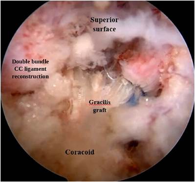
Fig 9. Viewing with the 4-mm 30 arthroscope from the midaxillary portal, the gracilis graft is reinforced with the FiberTape (Arthrex) passed through the coracoid tunnel and the 2 clavicle tunnels in a V fashion re-creating the coracoclavicular ligaments, with the patient in a semi-beachchair position for the right shoulder. The reconstruction of both coracoclavicular ligaments using the V configuration helps in re-creating the vertical stability of the acromioclavicular joint.
the coracoid have been performed to reduce the fracture risk theoretically. Increased risk of clavicular tunnel widening and fractures have been seen in the group treated with looping of graft around the coracoid when compared with the tunnel group. Patients with a looped graft around the coracoid may experience significant scapular motions, which can resulted in the abovementioned complications at higher rates.15 Drilling of the coracoid with the drill sleeve in a center- center or medial-center tunnel configuration has a high peak load to failure16 and coracoid drilling using conventional drilling results in eccentric drilling, producing stress risers and fractures.17-19
Our technique involves reconstruction of both coracoclavicular and acromioclavicular ligaments, providing both vertical and horizontal stability to the
Fig 11. Final surgical picture showing the secured graft restoring the horizontal and the vertical stability of the acromioclavicular joint as the coracoclavicular and the acromioclavicular ligament reconstruction is done, with the patient in a semi-beach-chair position for the right shoulder.
Fig 12. Viewing with the 4-mm 30 arthroscope from the midaxillary portal, the arthroscopic image shows the wellseated dog button under the coracoid process, with the patient in a semi-beach-chair position for the right shoulder. The dog button holding the graft under the coracoid and the tension on the graft are noted.
Fig 13. The animated figure showing the acromioclavicular ligament reconstruction and the surgical construct using the gracilis autograft reinforced with the FiberTape (Arthrex) and the dog button. The V configuration of the graft showing the reconstruction of the coracoclavicular ligament and acromioclavicular ligament is demonstrated.
acromioclavicular joint, restoring native physiological properties of the AC joint.20-23 An advantage of our technique is that the addition of a FiberTape internal brace along with the graft and the dog button prevents the loss of reduction due to creep of the soft tissue graft.19,24 The usage of a dog button gives good anchorage of the graft and also equal stress distribution to the coracoid. The double-stranded configuration of the graft provides anatomic double-bundle reconstruction of the coracoclavicular ligaments (Tables 1, 2).
Disclosures
All authors (A.V.N., P.K.M., A.J., P.K., P.S.K., M.R., S.T.) declare that they have no known competing
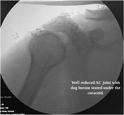
Fig 14. Intraoperative fluoroscopic image demonstrating well-reduced acromioclavicular joint with the dog button seated flush under the coracoid process.
Table 1. Tips and Tricks of the Technique
Tips
1.Clearing the medial border of the coracoid involving the release of pectoralis minor tendon ensures adequate visualization of the coracoid for tunnel drilling.
2.Open exposure of the acromioclavicular joint and removal of the scar tissue along with the distal end of the clavicle aids in obtaining good reduction of the joint.
3.Drilling of a coracoid tunnel using a sleeve from the superior surface ensures center-center tunnel position in the coracoid, reducing the risk of fracture.
4.Passage of graft through the coracoid tunnels provides good healing rates of the graft with no risk of graft displacement.
Tricks
1.Graft reinforced with FiberTape (Arthrex) reduces the incidence of graft stretching, leading to loss of reduction.
2.Trimming the graft to keep the graft diameter to 5 mm will help in easy passage of the graft and also to avoid a wider tunnel in the coracoid.
3.Use of 2 tunnels in the clavicle provides anatomic reconstruction of the coracoclavicular ligaments.
4.The reduction technique using a bone spike levering against the acromion aids in obtaining good reduction of the joint.
Table 2. Pitfalls of the Technique
1.Caution should be needed when using the trans-pectoralis major portal due to proximity of neurovascular structures.
2.Release of pectoralis minor tendon attachment has future risk of scapular dyskinesis.
3.Coracoid tunnel diameter more than 6 mm has risk of fracture of the coracoid.
4.Shuttling of the double-stranded graft through the coracoid tunnel should be done with care as there is a risk of graft bulging up during passage.
financial interests or personal relationships that could have appeared to influence the work reported in this paper.
References
- Pallis M, Cameron KL, Svoboda SJ, Owens BD. Epidemiology of acromioclavicular joint injury in young athletes. Am J Sports Med 2012;40:2072-2077.
- Hibberd EE, Kerr ZY, Roos KG, Djoko A, Dompier TP. Epidemiology of acromioclavicular joint sprains in 25 National Collegiate Athletic association sports: 2009-2010 to 2014-2015 academic years. Am J Sports Med 2016;44: 2667-2674.
- Fraser-Moodie JA, Shortt NL, Robinson CM. Injuries to the acromioclavicular joint. J Bone Joint Surg Br 2008;90: 697-707.
- Beitzel K, Mazzocca AD, Bak K, et al. ISAKOS upper extremity committee consensus statement on the need for diversification of the Rockwood classification for acromioclavicular joint injuries. Arthroscopy 2014;30:271-278.
- Phadke A, Bakti N, Bawale R, Singh B. Current concepts in management of ACJ injuries. J Clin Orthop Trauma 2019;10:480-485.
- Chang HM, Wang CH, Hsu KL, et al. Does WeavereDunn procedure have a role in chronic acromioclavicular dislocations? A meta-analysis. J Orthop Surg 2022;17:95.
- Gowd AK, Liu JN, Cabarcas BC, et al. Current concepts in the operative management of acromioclavicular dislocations: A systematic review and meta-analysis of operative techniques. Am J Sports Med 2019;47:2745-2758.
- Lamplot JD, Shah SS, Chan JM, et al. Arthroscopicassisted coracoclavicular ligament reconstruction: clinical outcomes and return to activity at mean 6-year follow-up. Arthroscopy 2021;37:1086-1095.e1.
- Ruiz Ibán MA, Moreno Romero MS, Diaz Heredia J, Ruiz Díaz R, Muriel A, López-Alcalde J. The prevalence of intraarticular associated lesions after acute acromioclavicular joint injuries is 20%. A systematic review and meta-analysis. Knee Surg Sports Traumatol Arthrosc 2021;29:2024-2038.
- Clavert P, Meyer A, Boyer P, et al. Complication rates and types of failure after arthroscopic acute acromioclavicular dislocation fixation. Prospective multicenter study of 116 cases. Orthop Traumatol Surg Res 2015;101:S313-S316
- Kimmeyer M, Hees T, Buijze GA, Lafosse L, Lafosse T. High vertical and horizontal stability at short-term followup after an all-endoscopic double cerclage endobutton technique for acute acromioclavicular joint separations. Arthroscopy published online March 26, 2024.
- Pauly S, Kraus N, Greiner S, Scheibel M. Prevalence and pattern of glenohumeral injuries among acute high-grade acromioclavicular joint instabilities. J Shoulder Elbow Surg 2013;22:760-766.
- Michlitsch MG, Adamson GJ, Pink M, Estess A, Shankwiler JA, Lee TQ. Biomechanical comparison of a modified Weaver-Dunn and a free-tissue graft reconstruction of the acromioclavicular joint complex. Am J Sports Med 2010;38:1196-1203.
- Park I, Lee S, Shin SJ. Arthroscopic-assisted doublebundle coracoclavicular ligament reconstruction using cortical fixation buttons with suture tape provides superior vertical stability than the single-bundle reconstruction for acute acromioclavicular joint dislocation. Arthroscopy 2023;39:213-221.
- Jeon N, Choi NH, Ha JH, Kim M, Lim TK. Clavicular tunnel complications after coracoclavicular reconstruction in acute acromioclavicular dislocation: Coracoid loop versus coracoid tunnel fixation. Clin Orthop Surg 2022;14: 128-135.
- Ferreira JV, Chowaniec D, Obopilwe E, Nowak MD, Arciero RA, Mazzocca AD. Biomechanical evaluation of effect of coracoid tunnel placement on load to failure of fixation during repair of acromioclavicular joint dislocations. Arthroscopy 2012;28:1230-1236.
- Hohmann E, Oduoye S, Myburgh J, Zyl R van, Keough N. Transcoracoid drilling for coracoclavicular ligament reconstructions in patients with acromioclavicular joint dislocations result in eccentric tunnels. Arthrosc Sports Med Rehabil 2023;5:e489-e494.
- Olivos-Meza A, Rodríguez-Argueta ME, Suarez-Ahedo C, Jiménez-Aroche CA, Pérez-Jiménez FJ, RodríguezSantamaria I. Free hand drilling technique to enhance central position of tunnels in arthroscopic acromioclavicular joint fixation using TightRope system. Arthrosc Tech 2021;10:e1829-e1837.
- Bellmann F, Eckl L, Vetter P, Audigé L, Scheibel M. Clavicular tunnel widening after acromioclavicular stabilization shows implant-dependent correlation with postoperative loss of reduction. Arthroscopy 2023;39: 2273-2280.
- Berthold DP, Muench LN, Dyrna F, et al. Current concepts in acromioclavicular joint (AC) instabilityda proposed treatment algorithm for acute and chronic AC-joint surgery. BMC Musculoskelet Disord 2022;23:1078.
- Cerciello S, Corona K, Morris BJ, et al. Hybrid coracoclavicular and acromioclavicular reconstruction in chronic acromioclavicular joint dislocations yields good functional and radiographic results. Knee Surg Sports Traumatol Arthrosc 2022;30:2084-2091.
- Lädermann A, Denard PJ, Collin P, Cau JBC, Van Rooij F, Piotton S. Early and delayed acromioclavicular joint reconstruction provide equivalent outcomes. J Shoulder Elbow Surg 2021;30:635-640.
- Peebles LA, Aman ZS, Kraeutler MJ, Mulcahey MK.
Qualitative and quantitative anatomic descriptions of the coracoclavicular and acromioclavicular ligaments: A systematic review. Arthrosc Sports Med Rehabil 2022;4: e1545-e1555.