Written by: Editorial Team of Dr Ayyappan V Nair
Medically Reviewed by: Dr. Ayyappan V. Nair

Senior Consultant – Shoulder Surgery, Arthroscopy and Sports Trauma

Manipal Hospitals, Bangalore
Last Reviewed: 8th August 2026

Anatomy and Biomechanics

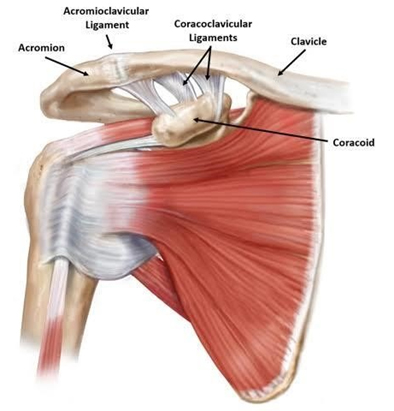
  • The Acromioclavicular (AC) ligament functions as a horizontal stabilizer, resisting 90% of anteroposterior translation and joint distraction.
  • The posterior and superior AC capsular ligaments provide the greatest degree of stability.
  • The Coracoclavicular (CC) ligaments act as vertical stabilizers and consist of two main components: the conoid and trapezoid ligaments.
  • The conoid ligament attaches posteriorly and medially on the clavicle, serving as the primary restraint to superior translation, and is the second ligament to fail after the AC ligament.
  • The trapezoid ligament attaches anteriorly and laterally on the clavicle, resisting 75% of AC joint compression.
  • Passive movements occur in coordination with the scapula when abducted beyond 90 degrees.
AC Joint Injury

Mechanism of Injury

Direct Mechanism
  • Most common cause, accounting for about 90% of AC joint separations. Typically results from a direct impact to the superior or lateral acromion (like a fall or tackle) while the arm is adducted.
  • The “Strut Effect”: the clavicle remains anchored anatomically while the force drives the acromion and scapula downward.
  • Sequential Failure: AC ligaments and capsule fail first, followed by CC ligaments (conoid fails before trapezoid), and finally the deltotrapezial fascia in high-grade injuries.
Indirect Mechanism
  • Less common, usually resulting from a fall on an outstretched hand (FOOSH) or the point of the elbow.
  • Axial load drives the humeral head upward into the acromion, selectively stressing the superior AC joint capsule and ligaments.
  • CC ligaments are usually spared because the acromion is pushed toward the clavicle, resulting exclusively in low-grade injuries.
Diagnosis and Clinical Evaluation
  • Clinical Presentation: Tenderness over the lateral end of the clavicle, swelling, deformity, and assessing joint reducibility.
  • Special Tests: O’Brien’s active compression test, Paxinos sign (cross-body adduction test), resisted extension test, and thorough SC joint/neurological exams.
  • Imaging: AP view (screening), Stress views, Zanca view (highly specific for AC joint), Axillary view (AP displacement), and Alexander view (differentiates Type IIIA and IIIB injuries).
Classification and Treatment Guidelines (Rockwood Classification)
Acromioclavicular (AC) Joint Injuries

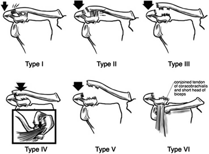
The ISAKOS Upper Extremity Committee subclassified Type III injuries into IIIA and IIIB based entirely on horizontal stability and scapular kinematics.

Type

AC / CC Status

Clinical Presentation

Radiographic Findings

Management

Type I

AC Sprain, CC Normal

AC tenderness, no instability

Normal

Conservative (Sling)

Type II

AC Torn, CC Sprain

AC horizontal instability

AC disrupted, CC distance < 25% increased

Conservative (Sling)

Type III

AC Torn, CC Torn

See IIIA and IIIB below

AC disrupted, CC distance 25-100% increased

Controversial

Type IIIA

Torn / Torn

AC vertical instability, no horizontal instability

N/A

Conservative

Type IIIB

Torn / Torn

AC vertical and horizontal instability

N/A

Surgical

Type IV

AC Torn, CC Torn

Skin tenting, posterior fullness

Lateral clavicle displaced posterior

Surgical

Type V

AC Torn, CC Torn

Severe shoulder droop unresponsive to shrug

Increased CC distance > 100%

Surgical

Type VI

AC Torn, CC Torn

Associated injuries, paresthesias (Rare)

Inferior dislocation of lateral clavicle

Surgical

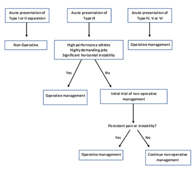
Note: Acute Type III presentations may require operative management for high-performance athletes or highly demanding jobs; otherwise, conservative management is recommended.

Management Protocols

Conservative Management Phases

  • Phase I (Weeks 0–2): Analgesia, controlling inflammation, preventing stiffness. Gentle active/active-assisted ROM, submaximal isometrics, weight-bearing < 500g. Sling discarded as rapidly as symptoms allow.
  • Phase II (Weeks 2–4): Restore full symmetric ROM, progressive load tolerance (weight-bearing < 5kg). Isotonic contractions for periscapular muscles; avoid heavy AC joint loading.
  • Phase III (Weeks 4–6): Proprioception and dynamic joint control using open-chain strengthening and PNF exercises. Weight-bearing restrictions removed.
  • Phase IV (Weeks 6–12+): Unrestricted functional participation, sport/occupation-specific training (kinetic chain/deceleration focus for throwing athletes).

Surgical Management Techniques

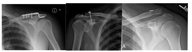
  • Fixation Across AC Joint: Phimester technique (K-wires) and Hook Plate.
  • Fixation Between Coracoid and Clavicle: Bosworth technique (screw fixation) or Tightrope device.
Acromioclavicular Ligament repair
  • Ligament Procedures: Ligament repair +/- augmentation, anatomical reconstruction, or distal clavicle excision.
  • Weaver-Dunn Procedure: Distal clavicle excision, transfer of CA ligament to distal clavicle, CC ligament and deltotrapezial fascia repair (AC joint not addressed).

OUR WORK

  • Arthroscopic Techniques: Single-button fixation over the clavicle using a FiberTape cinch loop through the coracoid for acute injuries.

“V” configuration anatomic reconstruction using double-bundled autogenous gracilis tendon graft reinforced with internal brace for chronic dislocations.

Reference:

https://doi.org/10.1016/j.eats.2024.103363
https://doi.org/10.1016/j.eats.2024.103154

For enquiries and online appointments, send a message to www.DrAyyappanVNair.com/contact

For informative videos related to Shoulder problems and their treatment options, Sports Injuries and other orthopedic conditions, visit our YouTube channel Bangalore Shoulder Institute – https://www.youtube.com/@BangaloreShoulderInstitute