

Arun Mundakkal, D.N.B. (Ortho), Jiss Joseph Panakkal, M.S. (Ortho),
Ayyappan V. Nair, D’Ortho, D.N.B. (Ortho), Siyad Ahammad, D’Ortho, D.N.B. (Ortho),
Jimmy Joseph Meleppuram, M.S. (Ortho), and
Prince Shanavas Khan, D’Ortho, M.S. (Ortho)
- Journal of Arthroscopy – 2023
LINK: https://www.arthroscopytechniques.org/article/S2212-6287(23)00317-1/fulltext
Arthroscopic repair of chronic retracted rotator cuff tears remains technically challenging, with high rates of incomplete healing and retear, especially in massive degenerative tears. The long head of the biceps tendon (LHBT) is highly vascular and rich in tenocytes and fibroblasts, making it a valuable biological augment. In massive degenerative tears where cuff tissue can be mobilized onto the footprint without undue tension, LHBT augmentation provides structural support and improves healing potential.
We describe an arthroscopic technique involving extensive cuff release, footprint preparation, biceps tenotomy, and double‑row repair augmented with the LHBT. The tenotomized biceps tendon is passed through the rotator cuff defect and secured to the cuff tissue and footprint using multiple double‑loaded suture anchors, including a crisscross medial‑lateral configuration. This approach improves tendon fixation, enhances footprint coverage, and provides a biologically active scaffold to support cuff healing. Indicated for massive retracted tears with adequate excursion after release, LHBT augmentation avoids the need for allograft or autograft harvest, reducing morbidity and cost. Early outcomes demonstrate improved tendon integrity and functional recovery. This technique offers a reproducible method to address poor‑quality massive cuff tears while preserving native tissue and enhancing biological healing.
Massive rotator cuff tears, when left untreated, tendon (LHBT) was later used for SCR instead of the progress with deterioration of the cuff autologous fascia lata graft or dermal allograft.3,4
| morphology and become irreparable. Many surgical methods have been described for massive irreparable rotator cuff tears, which include arthroscopic debridement with or without biceps tenotomy, partial repair of rotator cuff, tendon transfers, and superior capsular reconstruction (SCR).1 Superior capsular reconstruction restores the restraining effect of the superior joint capsule and balanced force couple in massive irreparable rotator cuff tears.2 The long head of the biceps | A lot of confusion exists regarding the difference between biceps augmented rotator cuff repair and a reverse biceps tenodesis (Biceps SCR). SCR using the LHBT is done by fixing the biceps tendon from the superior labrum onto the rotator cuff footprint, creating a reverse biceps tenodesis effect that leads to a downward force on the humeral head due to the rerouted LHBT and the space-occupying effect of the SCR. It is indicated in massive irreparable rotator cuff tears with an intact biceps tendon.5 Biceps augmented rotator cuff repair is indicated in extensively released retracted |
degenerate rotator cuff tears with an adequate excursion onto the rotator cuff footprint without undue tension. The biceps tendon provides a local autologous source of collagen graft that acts as an augment.6
In this Technical Note, we describe arthroscopic rotator cuff repair augmented with a LHBT in a massive rotator cuff tear with poor tissue quality. Surgical Technique
Surgery is performed under general anesthesia and interscalene-block anesthesia. The patient can be positioned in either the beach-chair or lateral decubitus position. We position the patient in a lateral position (Shoulder Suspension System; Arthrex, Naples, Florida). After sterile preparation and draping, bony landmarks are outlined.
e2 A. MUNDAKKAL ET AL.
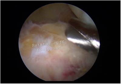
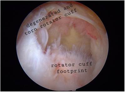
Fig 1. Patient in the lateral decubitus position. Diagnostic arthroscopy of right shoulder through posterior working portal. Through the anterior working portal, the extensive release of the torn rotator cuff is done to obtain excursion of the rotator cuff onto the footprint without tissue tension.
A standard posterior portal is used to perform a routine diagnostic arthroscopy of the glenohumeral joint. The degree of a rotator cuff tear, the lesions of the biceps tendon, and other intraarticular pathologies are evaluated. The arthroscope is then withdrawn and the arthroscope sheath is redirected into the subacromial space. A lateral portal is then made. A subacromial bursectomy is done. Acromioplasty may be done in indicated patients. Viewing through the lateral portal, the tear size is measured in the anterior-posterior direction using a calibrated probe through the posterior portal. Based on the size of the tear, degree of retraction, mobility, tissue thickness, and quality, reparability of the torn rotator cuff is assessed.
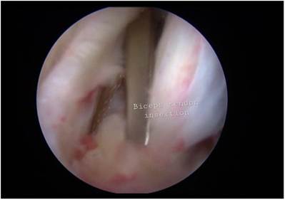
Fig 2. Patient in the lateral decubitus position. Viewing through the posterior portal. Through the anterior portal, the biceps tenotomy is done at the insertion.
Fig 3. Patient in the lateral decubitus position. Viewing through the posterior portal. Through the anterior portal, the rotator cuff footprint is debrided using a shaver.
The retracted torn rotator cuff is extensively released using a radiofrequency ablator. The decision to perform a biceps augmented rotator cuff repair or reverse biceps tenodesis depends on the remaining cuff tissue quality and mobility of the cuff after release. If footprint repair is not possible after adequate release and medialization of the footprint, we prefer to perform a reverse biceps tenodesis or SCR. If the cuff tissue is mobile enough for a footprint repair, the remaining tendon tissue quality is not good, and the suture limbs are in danger of a cutthrough, we perform rotator cuff repair augmented with biceps tendon (Fig 1).
The arthroscope is inserted into the glenohumeral joint. Using arthroscopic scissors or a radiofrequency probe, a biceps tenotomy is done at the insertion site close to the posterosuperior labrum (Fig 2). Another
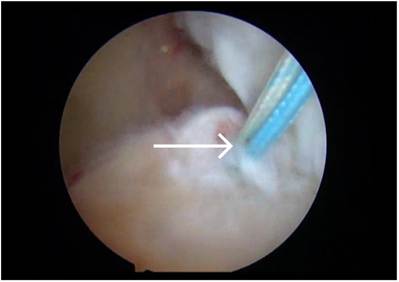
Fig 4. Patient in the lateral decubitus position. Viewing through the posterior portal. Through the anterior portal, a double-loaded anchor is inserted on the rotator cuff footprint anteriorly. The arrow mark shows the double-loaded anchor inserted.
| ROTATOR CUFF REPAIR WITH BICEPS AUGMENTATION e3 |
Fig 5. Patient in the lateral decubitus position. Viewing through the grand canyon view portal. The tenotomized biceps tendon is passed from the glenohumeral cavity into the subacromial space through the rotator cuff defect through the lateral portal.
portal is created posterior to the lateral portal midway between the posterior and lateral portal, known as the “grand canyon view portal,” which is employed as the viewing portal for further repair. The arthroscope is again inserted into the subacromial space. The rotator cuff footprint is debrided using a shaver (Fig 3). A double-loaded anchor (HEALICOIL; Smith & Nephew) is inserted on the rotator cuff footprint anteriorly (Fig 4). The 2 limbs of 1 suture of the double-loaded anchor are shuttled through the rotator cuff using a lasso suture passer. The tenotomized biceps tendon is passed from the glenohumeral cavity into the subacromial space through the rotator cuff defect through the lateral portal (Fig 5). The second suture of the
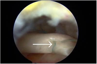
Fig 6. Patient in the lateral decubitus position. Viewing through the grand canyon view portal. Through the lateral portal, the second suture of the double-loaded anchor is retrieved through the substance of the biceps tendon using the lasso suture passer and tied using the lasso-loop technique. The first suture is shuttled through the rotator cuff using a lasso suture passer.
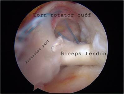
Fig 7. Patient in the lateral decubitus position. Viewing through the grand canyon view portal. Through the lateral portal, the proximal tip of the biceps tendon is tied to the posterior part of the torn rotator cuff with simple sutures.
double-loaded anchor is shuttled through the substance of the biceps tendon using the bird beak suture passer (Rhino suture passer; Arthrex) and tied using the lassoloop technique (Fig 6). The proximal tip of the biceps tendon is tied to the posterior part of the torn rotator cuff with simple sutures (Fig 7). A second doubleloaded anchor (HEALICOIL; Smith & Nephew) is inserted posteriorly at the rotator cuff footprint. The 4 sutures of the anchor are retrieved through the rotator cuff using the lasso suture passer and tied. The third double-loaded suture anchor (TwinFix Ti 5; Smith & Nephew) is placed between the first and second
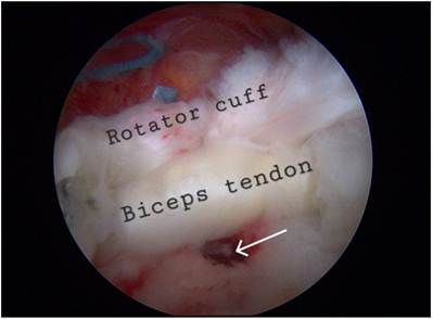
Fig 8. Patient in the lateral decubitus position. Viewing through the grand canyon view portal. Through the lateral portal, second and third double-loaded anchors are inserted at the footprint and all the sutures are retrieved through both the rotator cuff tissue and the biceps tendon using a lasso suture passer. The arrow shows the medial row anchor inserted.
| e4 A. MUNDAKKAL ET AL. |
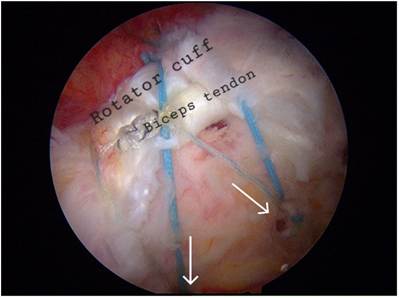
Fig 9. Patient in the lateral decubitus position. Viewing through the grand canyon view portal. Through the lateral portal, double-row rotator cuff repair is done using 2 lateral row anchors. The arrow shows the lateral row anchor.
anchors. The 4 limbs of the anchor are retrieved through both the rotator cuff and the biceps tendon proximal to the anchored part (Fig 8). Double-row repair of the rotator cuff is done with 2 lateral row anchors (SwiveLock; Arthrex) (Fig 9).
Postoperative Rehabilitation
In the immediate postoperative period, passive exercises, including pendulum, passive forward flexion, and external rotation exercises, are performed. Active range of motion is started only after 6 weeks postoperatively or after a full passive range of motion has been attained. At 6 weeks postoperatively, active-assisted exercises are initiated. Muscle-strengthening exercises are gradually introduced. For the first 6 months, activities and exercises that can cause stress to the shoulder are avoided.
Discussion
Arthroscopic repair of chronic retracted rotator cuff tears remains challenging to shoulder arthroscopy surgeons. Although 24% to 74% successful healing rates have been reported in massive rotator cuff tears, there is a high incidence of retears and incomplete healing.7 Table 1. Advantages and Disadvantages of Our Technique
| Pearls | Pitfalls |
| Structural support to the poor-quality rotator cuff tissue Enhance healing potential In contrast to biceps superior capsular reconstruction, biceps augmentation can be done even with an associated SLAP tear | Technically demanding Indicated in rotator cuff tears with an adequate excursion onto the cuff footprint without undue tension after releases Persistent pain at the biceps tendon groove |
With the recent technical advances, most of the massive rotator cuff tears are managed successfully. Due to the degenerative nature of the tears and their propensity to heal incompletely, grafting or augmentation of repair is desirable. The biceps tendon is highly vascular and a rich source of tenocytes and fibroblasts, which can promote biological healing.8 The LHBT provides an autologous source of collagen graft that can be performed arthroscopically without additional exposure or implants.6 In massive degenerate rotator cuff tears in which the rotator cuff tissue can be released and fixed onto the footprint without much tissue tension, the LHBT can act as an augment providing structural support to the poor-quality rotator cuff tissue and enhancing the healing process.
In massive irreparable rotator cuff tears, Mihata et al.9,10 proposed SCR using an autologous fascia lata graft to restore the restraining effect of the superior capsule and the balanced coupled force. The LHBT is used as an alternative to the fascia lata graft and dermal allograft in SCR, avoiding a second surgical site and donor site morbidity of the fascia lata graft and the high cost of a dermal allograft.3,4 Keeping the insertion at the superior glenoid and adjacent labrum intact, the LHBT is fixed at the rotator cuff footprint, creating a reverse tenodesis effect, which provides a downward force on the humeral head.
The decision between biceps augmented rotator cuff repair and reverse biceps tenodesis thus depends on the mobility of the cuff tissue after release and the tissue quality. If the rotator cuff tissue is mobile enough for a footprint repair but with poor tissue quality, the suture limbs through the rotator cuff tissue are in danger of a
cut-through. Augmenting double-row rotator cuff repair with the LHBT improves tissue grip by providing and enhancing healing potential by bringing new blood supply to the rotator cuff tissue.
The advantages and disadvantages of our technique are described in Table 1.
References
- Hermanowicz K, Góralczyk A, Malinowski K, Jancewicz P, Domzalski ME. Long head biceps tendon_ natural patch for massive irreparable rotator cuff tears. Arthrosc Tech 2018;7:e473-e478.
- Pauzenberger L, Hurley ET. Editorial commentary: Finally, something positive about the long head of the biceps tendon?! Shoulder superior capsular reconstruction. Arthroscopy 2018;34:2601-2603.
- Park MC, Itami Y, Lin CC, et al. Anterior cable reconstruction using the proximal biceps tendon for large rotator cuff defects limits superior migration and subacromial contact without inhibiting range of motion: A biomechanical analysis. Arthroscopy 2018;34:2590-2600.
| ROTATOR CUFF REPAIR WITH BICEPS AUGMENTATION e5 |
- Boutsiadis A, Chen S, Jiang C, Lenoir H, Delsol P, Barth J. Long head of the biceps as a suitable available local tissue autograft for superior capsular reconstruction: “The Chinese way.”. Arthrosc Tech 2017;6:e1559-e1566.
- Kim YS, Lee HJ, Park I, Sung GY, Kim DJ, Kim JH.
Arthroscopic in situ superior capsular reconstruction using the long head of the biceps tendon. Arthrosc Tech 2018;7:e97-e103.
Mihata T, McGarry MH, Pirolo JM, Kinoshita M, Lee TQ. Superior capsule reconstruction to restore superior stability in irreparable rotator cuff tears: A biomechanical cadaveric study. Am J Sports Med 2012;40:2248-2255.
Rhee SM, Youn SM, Park JH, Rhee YG. Biceps rerouting for semirigid large-to-massive rotator cuff tears. Arthroscopy 2021;37:2769-2779.
Cheppalli NS, Purudappa PP, Metikala S, Goel A, Singla A, Sambandam S. Using biceps tendon autograft as a patch in the treatment of massive irreparable rotator cuff tears improves patient-reported outcome scores: A systematic review. Arthrosc Sports Med Rehabil 2023;5:e529-e536.
Colbath G, Murray A, Siatkowski S, et al. Autograft long head biceps tendon can be used as a scaffold for biologically augmenting rotator cuff repairs. Arthroscopy 2022;38: 38-48.
Mihata T, Lee TQ, Watanabe C, et al. Clinical results of arthroscopic superior capsule reconstruction for irreparable rotator cuff tears. Arthroscopy 2013;29:459-470.