Written by: Editorial Team of Dr Ayyappan V Nair
Medically Reviewed by: Dr. Ayyappan V. Nair Senior Consultant – Shoulder Surgery, Arthroscopy and Sports Trauma
Manipal Hospitals, Bangalore
Last Reviewed: 14th August 2026
They are the high-energy injuries, often accompanied by a cascade of multi-system trauma like rib fractures, pneumothorax, and spine injuries.
Dr Ayyappan V Nair, considered one of the best shoulder surgeons in India says that for decades, the orthopedic community treated the vast majority of these fractures with “benign neglect,” trusting the robust muscular envelope to forgive significant displacements. However, the paradigm is shifting. As our understanding of shoulder biomechanics improves, we are increasingly holding the scapula to the same reduction and fixation standards as other major articular joints.
Here is a look at how we currently approach, evaluate, and manage scapula fractures.
Basic Anatomy: The Dynamic Stabilizer
Scapula is the vital link between the axial skeleton and the upper extremity.
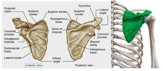
It serves as the attachment site for 18 different muscles, which create a thick soft-tissue envelope. This musculature provides tremendous blood supply for healing but also acts as a deforming force when the bone is fractured.
The structural integrity relies heavily on the Superior Shoulder Suspensory Complex (SSSC) – a bony and soft-tissue ring comprising the glenoid, coracoid, acromion, distal clavicle, and the connecting ligaments.
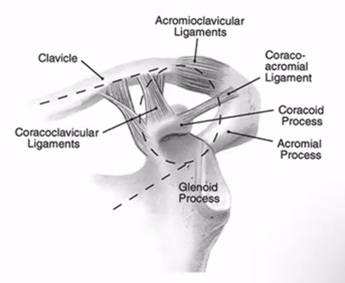
Disruptions to this ring (such as a “floating shoulder”) severely compromise the dynamic stability of the entire arm.
Classification
Classification is based on the location of the fracture and includes
- Scapular body fractures
- usually described based on anatomic location
- Scapular neck fractures
- look for associated AC joint separation or clavicle fracture
- Glenoid fractures
- Ideberg classification with Goss modification (below)
- Acromial fractures
- Kuhn classification
- Coracoid fractures
GLENOID FRACTURES
Type I: Glenoid rim (margin) avulsion/fracture (later subdivided into Type Ia anterior and Type Ib posterior by subsequent authors)
Type II: Fracture through the glenoid fossa exiting inferiorly/laterally
Type III: Oblique fracture exiting superiorly through the coracoid or superior shoulder girdle
Type IV: Transverse fracture exiting through the medial border of the scapula body
Type V: Combination of fracture patterns (e.g., Type II combined with Type IV)
Type VI category for severe, highly comminuted glenoid fractures was later added by Goss in subsequent modifications
Clinical Presentation
When a patient arrives with a suspected scapula fracture, the first priority is always clearing life-threatening associated injuries. Once stabilized, the orthopedic workup begins.
- History
- traumatic direct blow to shoulder or fall on outstretched arm
- scapula fracture may be missed or diagnosed late in presence of other distracting, traumatic injuries
- Symptoms -Diffuse, severe shoulder pain
- Physical exam
- Tendern on palpation
- Abnormal shoulder contour compared to contralateral site
Evaluation
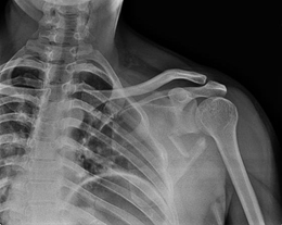
- Standard Radiographs: A true AP (Grashey), Scapular Y, and Axillary view are mandatory.
- 3D Computed Tomography (CT): This is the modern gold standard.
Plain films notoriously underestimate deformity.
A 3D CT reconstruction is essential for accurately measuring intra-articular step-offs, medialization, and the glenopolar angle (GPA), which directly dictate our surgical indications (Cole et al., 2012).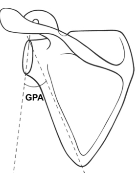
- GLENO POLAR ANGLE-
Normal:30-45 degrees
Management
Dr Ayyappan V Nair, Shoulder Surgeon says that over 90% of scapula fractures are minimally displaced and can still be managed conservatively with a brief period of sling immobilization followed by early, progressive range of motion to prevent adhesive capsulitis.
However, nonoperative management of severely displaced fractures consistently yields poor functional outcomes, including chronic pain, weakness, and altered shoulder kinematics (Cole & Schroder, 2021).
Current Surgical Indications:
While absolute consensus is still evolving, recent literature supports Open Reduction and Internal Fixation (ORIF) for the following thresholds:
- Intra-articular glenoid step-off or gap: ≥ 4 mm
- Glenopolar angle (GPA): ≤ 22°
- Lateral border offset (medialization): ≥ 20 mm (or ≥ 15 mm with significant angular deformity)
- Angular deformity: ≥ 45°
- Double disruptions of the SSSC with significant displacement.
Advanced Surgical Techniques
Approaches
While anterior glenoid rim fractures can be accessed via a standard deltopectoral approach, the vast majority of neck and body fractures require a posterior approach.
Traditionally, the classic Judet approach involved massive muscle reflection.
Today, we favor muscle-sparing modified posterior approaches that exploit internervous plane(the interval between the infraspinatus and teres minor). This minimizes soft-tissue devitalization and accelerates postoperative rehabilitation.
Reduction and Fixation
The overarching goal is to restore the lateral pillar. This maneuver is the linchpin of the operation; reducing the robust lateral border indirectly realigns the glenoid neck and restores the GPA.
- Implants:. Modern fixation relies on low-profile 2.4-mm and 2.7-mm locking and reconstruction plates. These are highly contourable to the complex, non-linear anatomy of the scapular spine and lateral border.
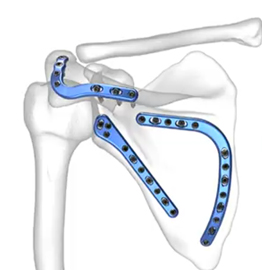
- Construct: A biomechanically stable construct usually involves plating the lateral pillar and extending fixation along the scapular spine or medial border to neutralize rotational forces.
For enquiries and online appointments, send a message to www.DrAyyappanVNair.com/contact
For informative videos related to Shoulder problems and their treatment options, Sports Injuries and other orthopedic conditions, visit our YouTube channel Bangalore Shoulder Institute – https://www.youtube.com/@BangaloreShoulderInstitute
References
Pires RE, Giordano V, de Souza FSM, Labronici PJ. Current challenges and controversies in the management of scapular fractures: a review. Patient Saf Surg. 2021 Jan 6;15(1):6. doi: 10.1186/s13037-020-00281-3. PMID: 33407725; PMCID: PMC7789406.
Cole, P. A., Gauger, E. M., & Schroder, L. K. (2012). Management of scapular fractures. Journal of the American Academy of Orthopaedic Surgeons, 20(3), 130–141. https://doi.org/10.5435/jaaos-20-03-130
Cole, P. A., & Schroder, L. K. (2021). Scapula Fractures. In Evidence-Based Orthopedics (pp. 457–461). Wiley. https://doi.org/10.1002/9781119413936.ch76