

Ayyappan V. Nair, D.Ortho, D.N.B., M.N.A.M.S., Pramod Kumar Mohan, M.S., Ortho.,
Pavan Krishna, M.S., Ortho., Ajit Jangale, M.S., M.R.C.S., Syed Kareemulla, M.S., Ortho., Naresh Goud, D. Ortho., D.N.B. Ortho., and Sreejith Thampy, M.S., Ortho.
- Arthroscopy Techniques – 2023
LINK: https://www.arthroscopytechniques.org/article/S2212-6287(23)00264-5/fulltext
Intraoperative vertical coracoid graft fractures are a recognized complication of the Latarjet procedure, often requiring additional iliac crest bone grafting, which increases patient morbidity. These fractures are attributed to excessive screw tightening, poor bone quality, smaller graft size, and are more common in females. This technical note describes a novel all‑arthroscopic double‑pulley salvage technique to address vertically split coracoid graft fractures without additional graft harvesting.
The technique uses two double‑loaded all‑suture anchors placed medially and laterally to the graft. Sutures are arranged in a double‑pulley configuration, providing controlled compression and stability to the fractured fragment while maintaining the benefits of the Latarjet procedure. This method allows the fracture to heal without removing the original screws, minimizing disruption to fixation.
The technique is reproducible, uses standard arthroscopic portals, and avoids the morbidity associated with iliac crest graft harvesting. Early results in two cases demonstrated good graft integration at three months and satisfactory clinical outcomes at one year. This salvage approach provides a viable alternative to traditional management of vertical coracoid graft fractures, reducing operative time and complications while preserving graft stability. The double‑pulley method is a promising adjunct for surgeons performing arthroscopic Latarjet procedures.
| A |
rthroscopic Latarjet procedure is the preferred literature and can be grouped as intraoperative, method of bony stabilization in recurrent hardware-related, graft-related, and neurovascular
6-9
| shoulder dislocation with certain criteria. Ideal candidates for the Latarjet treatment include patients with recurrent dislocation, glenoid bone loss higher than 15%, severe soft tissue insufficiency, failed Bankart repair, contact athlete dislocation, and ISIS greater than 4.1 The long-term outcomes following the Latarjet procedure are good to excellent with comparable results with an open and arthroscopic approach.2-4 However, the arthroscopic Latarjet procedure has the benefit of addressing additional intra-articular pathologies, such as SLAP lesions, and performing a remplissage for an off-track Hill-Sachs lesion. It also has the advantage of less postoperative pain with an earlier recovery, less bleeding, and slightly fewer instances of instability.5 Complications of the Latarjet procedure have been described in the | complications. Intraoperative graft fractures have been described, and this technical note aims to demonstrate an arthroscopic salvage procedure of intraoperative vertically split coracoid graft fractures (Video 1). In this technical note, we use a doublepulley configuration of suture tightening with 2 double-loaded, all-suture anchors and salvage of the vertically split coracoid graft are described. Two double-loaded, all-suture anchors (Stryker) are placed on either side of the vertically split coracoid graft and each suture threads are tied over the graft in doublepulley configuration, enabling graft compression and stability. Surgical Technique (With Video illustration) |
Patient Positioning and Portal Placement
| From the Bangalore Shoulder Institute, Bangalore, India. Received March 22, 2023; accepted August 15, 2023. Address correspondence to Pramod Kumar Mohan, M.S., Ortho, 40,36th Cross, 23rd Main Road, 4th T Block East, Jayanagar, Bangalore, 560041, Karnataka, India. E-mail: pramod.stanley@gmail.com 2023 THE AUTHORS. Published by Elsevier Inc. on behalf of the Arthroscopy Association of North America. This is an open access article under the CC BY-NC-ND license (http://creativecommons.org/licenses/by-nc-nd/ 4.0/). 2212-6287/23448 https://doi.org/10.1016/j.eats.2023.08.013 |
Under general anesthesia and an interscalene block, the patient is positioned in the beach chair position. The arm is prepped and draped, held by traction cable with the shoulder in 60 anterior elevation, 10 shoulder abduction, and neutral rotation (Fig 1).
Portal Placement
Surgical Steps
The arthroscopic Latarjet procedure, as described by
Lafosse, was performed. The coracoid graft was
Arthroscopy Techniques, Vol 13, No 1 (January), 2024: 102808 e1
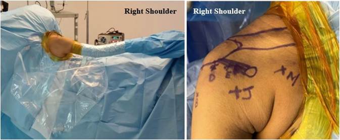
Fig 1. Patient positioning and portal placement. The patient is positioned in the beach chair position for the arthroscopic Latarjet
procedure, and the right shoulder is prepped and draped.
prepared, osteotomized, and transferred through the subscapularis split. The coracoid graft fixation was carried out with two cannulated metal screws (DepuyMitek, Chennai, India) and tightening of the screws was performed. While the tightening of graft was performed, we encountered an iatrogenic vertical split fracture of the coracoid graft, extending until the conjoint tendon insertion (Fig 2). The vertically split coracoid graft was salvaged with the arthroscopic technique, as described below.
Double-Loaded, All-Suture Anchor Placement
Using 4-mm 30 arthroscope, viewing from the mid axillary portal, two double-loaded, all-suture anchors (1.7 mm, Stryker) are placed. The first double-loaded anchor is placed on the medial scapular neck at the level of the coracoid graft, and the second doubleloaded anchor is inserted on the glenoid face at the same level as the first anchor. The anchors are inserted from the trans-pectoralis portal, we use a clear cannula (the anchor sheath is cut to the appropriate length for the anchor insertion) for anchor insertion. The suture threads of each anchor are identified as tiger blue and tiger black (Fig 3).
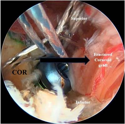
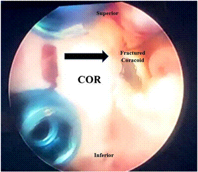
Fig 2. Viewing from the mid-axillary portal with a 4-mm 30 arthroscope in the right shoulder, the vertically split coracoid graft is visualized. The split coracoid graft is found secured with the glenoid bed using 2 titanium metal screws. COR, coracoid.
Fig 3. Viewing from the mid-axillary portal with a 4-mm 30 arthroscope in the right shoulder, the vertically split coracoid graft is visualized along with the 2 double-loaded, all-suture anchors on either side of the coracoid graft. One double-loaded, all-suture anchor is placed medially, and another anchor is placed laterally to the graft on the glenoid. COR, coracoid graft.
Suture Management and Placement of First Pulley
Viewing from the mid-axillary portal, the suture threads of each anchor are identified as tiger blue and tiger black. Initially, all the tiger blue suture threads from both the anchors and one tiger black suture thread from each anchor are parked outside the lateral portal. The remaining tiger black threads are retrieved from the trans-pectoralis portal making sure of no soft tissue entanglement. Both the threads are tied together with half-hitching knots from the trans-pectoralis portal through the clear cannula, 6 alternating half-hitching knots are placed. The remaining threads are cut after knotting. Then the other tiger black suture threads parked outside the lateral portal are retrieved from the trans-pectoralis portal, and tightening is done in a pulley fashion after loosening the inferior screw to get adequate compression of the fractured graft. The threads are secured with 6 alternating half-hitching knots and the remaining length of the 2 threads are taken over the graft and secured at the glenoid face at 5 o’clock position with a 2.7-mm push lock anchor (Arthrex, Naples FL) along with the sutures securing the capsulolabral tissue complex.
Second Pulley Placement
The single tiger blue suture threads from each anchor are retrieved through the trans-pectoralis portal. The suture threads are retrieved through the clear cannula, making sure there is no soft tissue window between the suture threads. Both threads are tied together with halfhitching knots from the trans-pectoralis portal through the clear cannula, 6 alternating half-hitching knots are placed. The remaining threads are cut after knotting. Then the other tiger blue suture threads parked outside the lateral portal are retrieved from the trans-pectoralis portal, and tightening is done in a pulley fashion. The ends of the suture threads are secured at the glenoid face at 4 o’clock position with 2.7-mm push lock anchors (Arthrex, Naples FL), along with the capsulolabral complex, making the graft extra-articular (Figs 4 and 5).
Final Arthroscopic Evaluation
Final arthroscopic evaluation done from the midaxillary portal showed adequate compression of the fractured coracoid graft with 2 metal screws. The double-pulley configuration provides adequate compression and sturdy fixation of the coracoid graft, allowing the graft to heal (Fig 6).
Postoperative Protocol
Postoperatively, the patient was immobilized in an arm sling. Shoulder shrugs, passive elbow mobilization, and hand grips were started from day 1 after surgery. Passive shoulder forward flexion and external rotation were started on the 10th day to achieve full passive
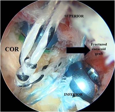
Fig 4. Viewing from the mid-axillary portal with a 4-mm 30 arthroscope in the right shoulder, the double-pulley configuration, using two double-loaded, all-suture anchors, is seen, along with the adequately compressed coracoid graft. COR, coracoid graft.
forward flexion and external rotation at the end of the 6 weeks. The patient was encouraged to perform computer work after suture removal on day 10 after surgery. Active shoulder movements and strengthening
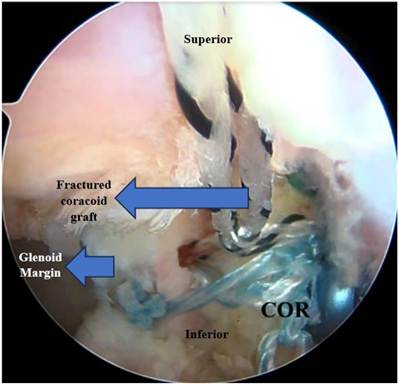
Fig 5. Viewing from the mid-axillary portal with a 4-mm 30 arthroscope in the right shoulder, fixation of the one end of the double-pulley along with capsulolabral tissue complex with 2.7-mm Push Lock anchor is visualized. COR, coracoid graft.
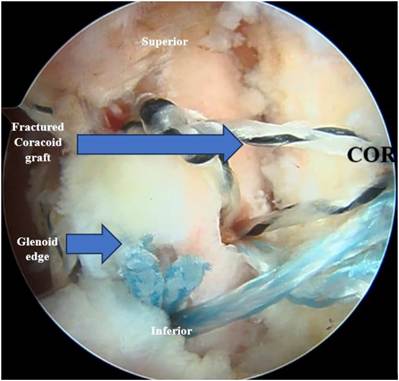
Fig 6. Viewing from the mid-axillary portal with a 4-mm 30 arthroscope in the right shoulder, 30 arthroscope, final construct with coracoid graft, capsulolabral tissue complex, and double-pulley configuration are secured with two 2.7-mm Push Lock is visualized. The 2.7-mm Push Lock is placed over the glenoid face, along with sutures and the capsulolabral tissue complex. COR, coracoid graft.
exercises were initiated at the end of 6 weeks and continued until the third month. The patient was allowed to return to daily activities in the sixth week, and sporting activities were allowed only after 6 months after the postoperative CT scan showed good bony union and remodelling.
Discussion
The learning curve in performing arthroscopic Latarjet was steep, which resulted in increased complication rates in the early learning period.7,10 Overall, complication rates following Latarjet procedure were estimated to be 17.3%.11 The advantages of arthroscopic Latarjet included less bleeding, less postoperative pain, less scarring, and early postoperative
mobilization.5,12,13
Intraoperative coracoid graft fractures were well documented in the literature with rates of 5.2e7% during the arthroscopic Latarjet procedure. Athwal et al. documented graft fractures, as the most common adverse effect following arthroscopic Latarjet and estimated their frequency to be 7%.14 Lafosse et al. documented graft fractures in 1% of their case series following arthroscopic Latarjet.15 The rates of coracoid graft fracture during open Latarjet were estimated to be 1.5%.16
Graft fractures during the Latarjet procedure were due to overtightening of the metallic screws, a smaller coracoid graft and poor quality of the bone.13,17 Athwal et al. proposed that good under-surface preparation of the coracoid graft can reduce the incidence of graft fractures. Maintaining 9-mm distance between the screw holes, using the two-finger technique for screw tightening, using top hats with metallic screws, and ensuring adequate graft length, adequate coracoid and glenoid preparation can reduce the graft fracture rates during the procedure.14
Depending on the fracture pattern and the stability of the graft after fixation, intraoperative graft fractures can be addressed. Transverse graft fractures may occur at the level of the beta screw (proximal screw), between
Table 1. Advantages, Pitfalls and Pearls of the Salvage
Technique for Coracoid Graft Fracture
Advantages
- The technique is relatively easy and reproducible for arthroscopy surgeons trained in arthroscopic Latarjet.
- The technique can also be used for graft salvage, while performing open Latarjet.
- The same portals used for arthroscopic Latarjet can be used forthis technique.
- Double-loaded, all-suture anchors are easier to place on bothsides of the coracoid graft due to the low profile of the anchors.
- The double-pulley method enables adequate compression of thefracture fragments.
Pitfalls
- Technically demanding for surgeons who are not trained in thearthroscopic Latarjet procedure
- Only vertical split fractures can be salvaged with this technique.
- Very osteoporotic coracoid is difficult to salvage using the method.
Pearls:
- Adequate visualization of the fractured graft is essential forperforming the technique.
- Cortical screws should be loosened to enable achieving adequate compression of the graft.
the beta and alpha screws, at the level of the alpha screw (distal screw), or at the joint tendon attachment. Depending on the location of the fracture, the fracture may be addressed. The flowchart outlines the management procedure to be followed in the event of transverse graft fractures (Fig 7).
Coracoid graft fractures that were not salvageable were treated with the Eden-Hybinette procedure, which requires iliac crest graft harvest. Although the procedure provided satisfactory outcomes, donor site morbidity should be considered. With our method, vertical split graft fractures that were previously treated with iliac crest bone grafting (Eden-Hybinette) can be salvaged. One advantage of our salvage technique is it can be performed arthroscopically and can also be used while performing open Latarjet. The technique was relatively easy and reproducible, ensuring dependable results. Kongmalai et al. proposed a salvage method for vertical graft fractures using 2 suture anchors on the medial glenoid neck during open Latarjet procedure.18 The advantages of our proposed technique are that using two double-loaded, all-suture anchors on either side of the graft in a pulley fashion provides adequate compression of the fractured graft, and it can be done in all-arthroscopic fashion (Table 1). We retained the metal screws instead of removing the screws; the screws were loosened a bit to achieve adequate graft compression and left in place (Fig 8).
Improper visualization of the graft while securing the knots following the double-pulley technique will result in graft loosening due to slackness of the knots. Coracoid graft fractures following arthroscopic Latarjet performed with cerclage tapes or cortical button are at risk of failure if double-pulley salvage technique is tried in such scenarios. Overcompression of the double pulley
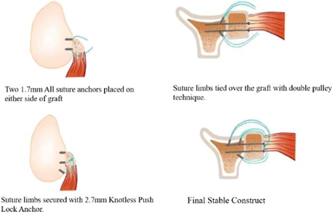
Fig 8. Animated figures showing the double-pulley salvage technique for vertically split coracoid graft in the right shoulder. The graft is salvaged with two double-loaded, all-suture anchors passed over the graft using the double-pulley fashion and securing the ends of the sutures with 2.7-mm Push Lock anchor.
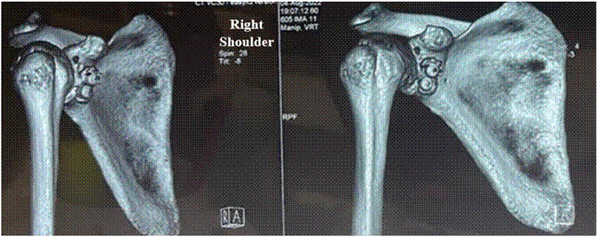
Fig 9. CT scans of the right shoulder at 3 months after Latarjet surgery showing good bony union of the coracoid graft. The coracoid graft is found to be incorporated with the glenoid, and there is no evidence of graft loosening or screw loosening.
Table 2. Risks and Limitations of the Procedure
1.All-suture anchors are preferred for double pulley due to the lowprofile nature of the anchors.
2.Osteoporosis can result in crushing of the graft when compression is applied with the double pulley.
3.Double-pulley technique of graft salvage can be done preferably following fractures encountered during arthroscopic screw Latarjet.
4.Improper visualization while securing the arthroscopic knots may result in slackness of the knots and subsequent graft loosening.
can cause crushing of the graft in cases where there is a very osteoporotic coracoid. Requirement of additional graft sources should be anticipated following graft fractures while performing the Latarjet procedure.
We have two cases of vertically split intraoperative coracoid graft fractures, which were salvaged with the proposed treatment method. We followed up the patients with CT scan at the third month to assess bony integration, as well as clinical follow-up for 1 year after surgery. We found good bony union in follow-up CT scans and good clinical outcomes following the graft salvage technique (Fig 9).
The all-arthroscopic salvage technique for the intraoperative vertical split of coracoid graft fracture is a simple, reproducible, and dependable option for coracoid graft salvage. Good preparation of the graft and avoiding overtightening of the screws can help avoid graft fracture. Being female, having osteoporotic grafts, and having smaller graft lengths were risk factors for graft fractures (Table 2).
Disclosure
The authors report no conflicts of interest in the authorship and publication of this article. Full ICMJE author disclosure forms are available for this article online, as supplementary material.
References
- Arner JW, Tanghe K, Shields T, et al. Shoulder Latarjet surgery shows wide variation in reported indications, techniques, perioperative treatment, and definition of outcomes, complications, and failure: A systematic review. Arthroscopy 2022;38:522-538.
- Wong SE, Friedman LGM, Garrigues GE. Arthroscopic
Latarjet: Indications, techniques, and results. Arthroscopy 2020;36:2044-2046.
- Chillemi C, Guerrisi M, Paglialunga C, Salate Santone F, Osimani M. Latarjet procedure for anterior shoulder instability: A 24-year follow-up study. Arch Orthop Trauma Surg 2021;141:189-196.
- Hurley ET, Ben Ari E, Lorentz NA, et al. Both open and arthroscopic Latarjet result in excellent outcomes and low recurrence rates for anterior shoulder instability. Arthrosc Sports Med Rehabil 2021;3:e1955-e1960.
- Getz CL, Joyce CD. Arthroscopic Latarjet for shoulder instability. Orthop Clin North Am 2020;51:373-381.
- Hurley ET, Schwartz LB, Mojica ES, et al. Short-term complications of the Latarjet procedure: A systematic review. J Shoulder Elbow Surg 2021;30:1693-1699.
- Bishai SK, Ball GRS, King C, et al. Arthroscopic Latarjet learning curve: Operating time decreases after 25 cases. Arthrosc Sports Med Rehabil 2023;5:e179-e184.
- Marjanovic B, Poberaj B, Kolar M, et al. 11% complications rate after Latarjet procedure at up to 14 years followup. Musculoskelet Surg 2022;106:227-237.
- Hendy BA, Padegimas EM, Kane L, et al. Early postoperative complications after Latarjet procedure: A singleinstitution experience over 10 years. J Shoulder Elbow Surg 2021;30:e300-e308.
- Bøe B, Støen RØ, Blich I, Moatshe G, Ludvigsen TC. Learning curve for arthroscopic shoulder Latarjet procedure shows shorter operating time and fewer complications with experience. Arthroscopy 2022;38:2391-2398.
- Buda M, D’Ambrosi R, Bellato E, et al. Failed Latarjet procedure: A systematic review of surgery revision options. J Orthop Traumatol 2021;22:1-16.
- Leuzinger J, Brzoska R, Métais P, et al. Learning curves in the arthroscopic Latarjet procedure: A multicenter analysis of the first 25 cases of 5 international surgeons. Arthroscopy 2019;35:2304-2311.
- Domos P, Lunini E, Walch G. Contraindications and complications of the Latarjet procedure. Shoulder Elbow 2018;10:15-24.
- Athwal GS, Meislin R, Getz C, Weinstein D, Favorito P. Short-term complications of the arthroscopic Latarjet procedure: A North American experience. Arthroscopy J 2016;32:1965-1970.
- DumontGD,FogertyS,RossoC,LafosseL.Thearthroscopic Latarjet procedure for anterior shoulder instability: 5-Year minimum follow-up. Am J Sports Med 2014;42:2560-2566.
- Griesser MJ, Harris JD, McCoy BW, et al. Complications and re-operations after Bristow-Latarjet shoulder stabilization: a systematic review. J Shoulder Elbow Surg 2013;22: 286-292.
- Mizuno N, Denard PJ, Raiss P, Melis B, Walch G. Longterm results of the Latarjet procedure for anterior instability of the shoulder. J Shoulder Elbow Surg 2014;23: 1691-1699.
- Kongmalai P, Levine WN. Double-row fixation of the transferred coracoid process: A rescue technique for intraoperative coracoid fracture during the Latarjet procedure. J Shoulder Elbow Surg 2019;28:e226-e231.