

Ayyappan V. Nair, D’Ortho, DNB Orthoa, Jiss Joseph Panakkal, MS Orthob, Pramod K. Mohan, MS Orthoa, Maythilisharan Rambhojan, MS Orthoa,
Sreejith Thampy J, MS Orthoa, Prince Shanavas Khan, D’Ortho, MS Orthoc,*
- JSES International – 2024
LINK: https://pubmed.ncbi.nlm.nih.gov/39280165/
This prospective study evaluated short-term outcomes of the arthroscopic Latarjet procedure using cerclage FiberTape fixation combined with remplissage and capsulolabral repair for recurrent anterior shoulder instability with glenoid bone loss. Ten male patients (mean age 28 ± 8.8 years) underwent surgery between 2020 and 2022 by a single fellowship-trained surgeon. Inclusion criteria included glenoid bone loss >15% or >10% with off-track Hill-Sachs lesions, failed Bankart repair, high instability severity index score, or contact athlete status. Functional outcomes were assessed pre- and postoperatively using VAS, ASES, Rowe, and Walch-Duplay scores, and graft position and healing were evaluated with CT at 3 months.
At a mean follow-up of 13.2 months, all patients showed significant improvements in pain and shoulder function. Mean postoperative ASES, Rowe, Walch-Duplay, and VAS scores were 95, 94.5, 87, and 1, respectively. CT scans revealed graft union in seven patients, with five flush-position grafts and two medial positions. Three patients (30%) demonstrated graft loosening and migration, but none required revision surgery or reported instability recurrence. No symptomatic hardware complications, infections, or neurovascular injuries occurred.
The study concludes that although cerclage FiberTape fixation avoids screw-related complications, it is associated with higher graft loosening rates compared to traditional screw fixation. However, functional outcomes remained excellent regardless of graft union status, suggesting the combined remplissage and capsulolabral repair contributed to stability. Long-term studies are needed to determine the durability of this technique.
Level of evidence: Level IV; Case Series;
Treatment Study
Background: The rates of implant-related complications are significant following the Latarjet procedure using metal screws in patients with recurrent shoulder dislocation and bone loss. The purpose of this study is to evaluate the short-term outcome following the arthroscopic Latarjet procedure using cerclage FiberTape (Arthrex, Naples, FL, USA) combined with remplissage and capsulolabral repair. It was hypothesized that performing the procedure with cerclage FiberTape would provide sturdy fixation, comparable to the conventional method of using metal screws, while averting hardware-related complications attributed to the latter in published literature.
Methods: A prospective study was performed in a single institution between 2020 and 2022, with all surgeries performed by a single fellowship-trained shoulder surgeon who has ample experience in performing arthroscopic screw Latarjet procedures. Patient demographics, number of dislocations before surgery, arm dominance, ligamentous laxity, type of sporting activity, Instability Severity Index Score, and percentage of bone loss on the glenoid and humeral sides were recorded. The patients were followed up with visual analog scale, American Shoulder and Elbow Surgeons score, Rowe score, and WalchDuplay score preoperatively and postoperatively. The coracoid graft position, healing, and remodeling were assessed with computed tomography scans at 3 months postoperatively. Minimum clinical followup was for a period of one year.
Results: Overall, 10 patients (all males, average age 28 ± 8.8 years) were operated on with an arthroscopic Latarjet procedure using cerclage FiberTape. The minimum follow-up period was 12 months, and the mean follow-up was 13.2 months. The median and individual visual analog scores during arm motion, American Shoulder and Elbow Surgeons scores, Rowe scores, and Walch-Duplay scores improved in the follow-up period. Computed tomography scans at 3 months showed flushed graft position in 5 patients, medial graft position in two patients, and three patients showed graft nonunion with migration. Out of 10 patients, seven had good graft union in follow-up scans. None of the patients required revision surgery. All three patients with graft nonunion were kept under follow-up beyond the study period for recurrence of instability.
Conclusion: Our study demonstrated that arthroscopic Latarjet using cerclage FiberTape fixation combined with remplissage and capsulolabral repair resulted in high rate of graft loosening and migration (30%). Nonetheless, patients in whom the coracoid graft had united, as well as those in whom it had not, all had good to excellent functional and clinical outcomes, no complications, and did not require any revision surgery.
© 2024 The Author(s). Published by Elsevier Inc. on behalf of American Shoulder and Elbow Surgeons.
This is an open access article under the CC BY-NC-ND license (http://creativecommons.org/licenses/bync-nd/4.0/).
46%.3 Complications from the use of metal screws include screw pull-out, loosening, breakage, intraarticular hardware, graft fracture, and osteolysis around screws.10 Some alternatives for graft fixation have been studied to prevent these complications. Buttress plates,8 bioabsorbable screws,2 cortical buttons,22 and cerclage FiberTape13 are examples of other methods of fixation mentioned in the literature that have been employed as substitutes to metal screws.
The purpose of this study was to analyze the position, rate of union, and osteolysis of the coracoid graft on computed tomography (CT) scan at 3 months, clinical and functional outcomes, as well as implant-related complications, at one year following the arthroscopic Latarjet procedure using cerclage FiberTape (Arthrex, Naples, FL, USA), combined with remplissage and capsulolabral repair. The authors hypothesized that the use of cerclage FiberTape can eliminate the screw-related complications while providing sturdy fixation of the graft, enabling good graft union, and also improving the clinical outcomes after the surgery.
Methods
This was a prospective study including consecutive patients who underwent arthroscopic Latarjet using Cerclage FiberTape combined with remplissage and capsulolabral repair operated by a single surgeon at a single institution between 2020 and 2022 with a minimum follow-up of 1 year. Patients with less than 1 year of follow-up and using an iliac crest graft were excluded from the study. Institutional review board approval was obtained for this study. The inclusion criteria included: 1) patients with glenoid bone loss >15% but <25%; 2) patients with glenoid bone loss >10% with off-track Hill-Sachs lesion; 3) dislocation in contact athletes; 4) failed Bankart repair; or 5) severe soft tissue insufficiency with ISI (Instability Severity Index) score >6. Patient demographics, number of dislocations before surgery, arm dominance, ligamentous laxity, type of sporting activity, ISI score, and percentage of bone loss on the glenoid and humeral sides were recorded.
The range of motion (expressed in degrees) was recorded preoperatively and after surgery. The forward flexion, abduction, external rotation by side (ER1), external rotation at 90-degree abduction (ER2), and internal rotation at 90-degree abduction were recorded before surgery and at the final follow-up. The functional outcomes were recorded with visual analog scale (VAS), American Shoulder and Elbow Surgeons (ASES) scores, Rowe scores, and Walch-Duplay scores preoperatively and after surgery at the final follow-up (at 12 weeks, 6 months, and one year postoperatively).
The graft position, graft union, and graft remodeling were assessed with CT scans at 3 months postsurgery. The graft position with respect to the anterior glenoid articular rim was assessed in axial CT slice at 50% of the glenoid height (distance between superior and inferior tubercles). It was classified as flush when perfectly aligned to the glenoid rim, medial, or lateral when more than a 5mm or 3mm step off was noted, respectively.4 In the vertical plane, the graft position was assessed at 50% of the glenoid height in a 3D en face view of the glenoid with the humeral head substituted. Presence of bony union and lysis was also determined. Patients who had nonunion of coracoid graft were followed up periodically beyond the study period for recurrence of instability.
Potential implant-related complications1,11,17 such as intraarticular penetration of implant/tunnel and graft fracture due to overtightening were assessed intraoperatively and on postoperative CT scans and patients were assessed for shoulder tenderness and pain for implant prominence and clinical signs of suprascapular nerve injury during the follow-up visits.
Statistical analysis was performed using non parametric test and Spearman rho tests. Non parametric test was employed to find the median of preoperative and postoperative clinical and functional scores. P value was determined by the Wilcoxon signed-rank test and the Spearman rho test was used to analyze the correlation of ISI scores, Beighton scores, and number of preoperative dislocations to the clinical and functional outcomes.
Preoperative assessment
The patients with recurrent anterior shoulder dislocation were assessed with 3D CT and magnetic resonance imaging. The amount of glenoid bone loss and the glenoid track were measured using the best-fit circle method from the “en face” view of the glenoid with humeral head suppression.21 The Hill-Sachs interval was measured from an axial CT scan and compared with the previously measured glenoid track to assess if the Hill-Sachs lesion was engaging or nonengaging.15
Surgical technique
Patient positioning and portal placement
The patient was positioned in a beach chair position under general anesthesia with an interscalene block. The arm was prepped and draped, held by traction cable, with the shoulder in 60-degree anterior elevation, 10-degree shoulder abduction, and neutral rotation. Standard Latarjet portals, as described by Lafosse were used.18 The surgeries were performed by the senior author using an all-arthroscopic Latarjet procedure. Diagnostic arthroscopy was performed, and associated pathologies were assessed through the posterior portal. The remplissage of the Hill-Sachs lesion was done in all patients with tightening of the remplissage sutures done as the final step after coracoid graft fixation. The coracoid preparation was done by skeletonizing the attached tissue (Fig. 1). The coracoacromial ligament was released from the coracoid attachment. The pectoralis minor tendon was released from the medial aspect of the coracoid from the M portal. Drill holes were made on the coracoid using the drill guide (DePuyMitek Raynham, MA, USA). The coracoid graft was harvested along with the conjoint tendon using the curved osteotome (DePuyMitek Raynham, MA, USA) through the H portal. The undersurface of the coracoid graft and the medial spike of the osteotomized graft were trimmed to achieve good bone contact while fixation to the glenoid (Fig. 2). The subscapularis split was made after passing a switching stick from the posterior portal. The subscapularis split was intended at the level of superior 2/3rd and inferior 1/3rd of the muscle. The split was carried out until the visualization of the glenoid neck and the glenoid bed was prepared with the burr at the intended site of coracoid fixation. The coracoid graft was captured using the positioning cannula (DePuy-Mitek Raynham, MA, USA) used for arthroscopic screw Latarjet, and the graft was transferred through the subscapularis split using the cannula. The position of the graft was confirmed with the switching stick passed flush to the glenoid face. The graft was secured with two beath pins with suture eye drilling from anterior to posterior direction through the positioning cannula, superior beta hole, and inferior alpha hole, exiting the posterior aspect of the shoulder. The Nitinol wire loop was passed through a beath pin in the beta hole from the anterior to posterior direction, exiting the posterior aspect of the shoulder. One end of the cerclage FiberTape (Arthrex, Naples, FL, USA) was transferred from the posterior to anterior direction, exiting the anterior surface of the shoulder through the nitinol wire loop. Another nitinol wire loop was passed through the beath pin in the alpha hole from the anterior to posterior direction, exiting the posterior aspect of the shoulder. The already transferred end of the cerclage FiberTape was passed through the nitinol wire loop from the anterior surface of the shoulder and
capsulolabral complex; SSC, subscapularis.
taken out through the posterior aspect of the shoulder. Both ends of the cerclage FiberTape were secured from the posterior aspect of the shoulder, and the looping of the cerclage FiberTape was done as per the protocol. The cerclage FiberTape was interconnected, and tensioning was done with the tensioning device to achieve a maximum tension of 80N, overtensioning may result in graft fractures. The final construct was visualized, ensuring proper bony contact between the coracoid and glenoid neck and also there was no slack in the cerclage FiberTape construct. The entire process of tightening of cerclage FiberTape was visualized from portal J. The capsulolabral tissue was anchored to the glenoid face with a 2.7-mm Arthrex push-lock, leaving the coracoid in an extracapsular position (Fig. 3). The tightening of the remplissage suture anchors was done as the final step. The modifications done were glenoid drill holes made using the positioning cannula with freehand technique instead of the glenoid drill jig (Video 1).
Rehabilitation protocol
The patient’s arm was immobilized with an arm sling for 6 weeks. Multidrug therapy and cryotherapy were used for pain control. Shoulder shrugs and assisted elbow flexion with hand grips were initiatedfromday 1. Passiveand active assisted forward flexion and external rotation were initiated from week 2 to attain 180 degrees of forward flexion and 45 degrees of external rotation at the end of week 6. Active shoulder movements and strengthening exercises were started in week 6 to attain a full range of motion and muscle power at the end of week 12. Contact sports were allowed after6 monthsafterensuring good graft union in follow-up CTscans. Regarding return to sports, a more cautious approach was employed for one patient with coracoid graft nonunion, who ultimately resumed contact sports (kabaddi) after 9 months.
Results
Demographics
Ten patients underwent an arthroscopic Latarjet procedure with cerclage FiberTape (all males) with an average age group of 28 ± 8.8 years (range 18-50). Six out of 10 patients were operated in their dominant shoulder. Three patients were involved in active contact sports, and one patient had a history of failed Bankart repair in the past. The average glenoid bone loss was 16 ± 4.0% (range 10-25%), and 6 patients had off-track Hill-Sachs lesions. Seven patients had glenoid defects 15% (range 15-25%), and 3 patients had 10% (range 10-14%) with associated ISI scores >6. The average follow-up period was 13.2 months. One patient had reached a follow-up of 24 months, and nine patients had a follow-up period of 12 months.
(Table I).
Pain scores
The pain scores during arm motion were recorded with VAS score. VAS scores were reduced from preoperatively mean 3.7 (range 0-6) to postoperatively mean 1 (range 0-2) at the final follow-up.
Clinical assessment
Results of range of motion assessment at one year follow-up are displayed in Table II. None of our patients had clinical evidence of neurological compromise. There were no complaints suggestive of symptomatic hardware.
Functional outcomes
At the final follow-up, the median and individual functional scores had significantly improved in all patients in both the union and nonunion groups. The mean ASES, Rowe, Walch-Duplay, and VAS scores were 95 (range 88-100), 94.5 (range 90-100), 87 (range 70-100), and 1 (range 0-2), respectively. None of our patients had any episodes of subluxation or instability.
Radiological assessment
- Graft position: Graft position was found flush in 5 patients, and 2 patients had a graft in the medial position. Three patients showed graft loosening and migration of the graft by more than 0.5 cm in follow-up CT. In the vertical plane, all the united coracoid grafts were found to be subequatorial.
- Graft healing: Seven patients showed good union of the coracoid healing in the final follow-up CT scan (Fig. 4, A and
B).
- Osteolysis: No osteolysis of the coracoid graft and no widening of the glenoid tunnels were noted.
- No coracoid graft fracture or intra-articular penetration ofthe graft tunnel was noted.
Patients who had achieved union of coracoid graft were referred to as being in “graft union group,” and those who had nonunion of coracoid graft were in “graft nonunion group.” (Table II). The mean postoperative ER1 and ER2 were significantly higher than the respective preoperative values (mean preoperative ER1 ¼ 54 vs. 68.5 postoperatively, mean preoperative ER2 ¼ 49 vs. 67 postoperatively). However, despite this amelioration, the mean postoperative ER1 and ER2 remained lower than the respective values of the opposite shoulder (mean ER1 76.5 and mean ER2 75).
Statistical analysis
A statistically significant improvement, defined by P value less than 0.05, was seen in all functional outcome scores (ASES, VAS, Walch-Duplay, and Rowe scores), ER1, ER2 and IR2 of the coracoid graft union group. Although significant improvements were seen in the graft nonunion group, reflecting those in the graft union group, the authors refrained from emphasizing a statistical comparison of these two groups bearing in mind the overall small sample size, compounded by the discrepancy in number of patients between the two groups. No statistically significant correlation was found between ISI score, Beighton score, and number of preoperative dislocations to the functional and clinical outcomes. (Table III). However, the authors express caution in their interpretation of this finding given the small sample size.
complex; GL, glenoid; SSC, subscapularis.
Table I
Patient demographics.
| Age, mean ± SD (range) | 28 ± 8.8 (18-50) |
| Sex, male/female | 10 males |
| Side, right/left Glenoid bone loss | 6 right/4 left |
| >20% | 2 |
| >15-20% | 5 |
| >10-14% | 3 |
| Bankart failure Glenoid tracking | 1 |
| Off-track | 6 |
| On-track | 4 |
SD, standard deviation.
Complications
Out of the ten patients, three showed coracoid graft loosening and migration in their 3rd monthly follow-up CT scans. The CT scans showed migrated graft (>0.5cm) with intact coracoid tunnels (Fig. 4, C and D). All three patients were kept under follow-up beyond the study period for recurrence of instability; they have had no symptoms of subjective instability. None have required any further interventions to date. There were no infection or neurovascular complications in our study. No symptoms related to symptomatic hardware were reported.
Discussion
In our study, we found out that Latarjet surgery using cerclage FiberTapes had high incidences of graft nonunion and migration in follow-up, without recurrence of instability or additional surgeries. The traditional Latarjet procedure using two metal screws was time-tested and effective in treating recurrent anterior instability.18,23 Long-term follow-up studies of the Latarjet procedure with metal screws demonstrated complication rates between 15% and 30%.3,1,11 The short-term follow-up of arthroscopic Latarjet also demonstrated 24% complication rates at 3 months.1
Implant-related complications following the Latarjet procedure were reported to be between 6.5% and 46%.3 Screw-related complications such as intra-articular screw prominence, screw breakage, screw bending, early arthritic changes of the humeral head, and soft tissue irritation (irritation of capsular tissue, subscapularis, or infraspinatus muscle) have been reported.4,11 Iatrogenic injury of the suprascapular nerve has been reported with anterior to posterior drilling and prominent screws.17 Long-term studies following the arthroscopic Latarjet procedure by Lafosse reported 12.5% revision surgeries for screw removal.6 Intraoperative graft fractures following metal screws were reported at around 7%, more common in young females with small coracoid grafts and excessive screw tightening.1 None of these complications were found in our study.
To reduce screw-related complications, other modes of fixation devices were tried by different authors. Bioabsorbable screws were used for graft fixation, but the long-term follow-up studies reported an increased rate of graft osteolysis and recurrent instability warranting revision surgeries.2 Mini-plate fixation of the coracoid graft demonstrated superior fixation strength over screw fixation.8,7 Chaudhary et al demonstrated satisfactory outcomes after mini-plate fixation,5 while mini-plate fixation needed extensive soft tissue dissection and caused soft tissue irritation. Suture button fixation demonstrated good outcomes with good union rates of bone block and also reduced revision surgeries when compared to metal screws.3 Hardy et al reported an increased risk of redislocation with suture button fixation compared to metal screws.13 Compared to rigid and metal implants, the potential benefits of using cerclage FiberTape were avoiding magnetic resonance
Table II
Clinical data, preoperative, and postoperative range of motion values and scores.
| Patient | Age (years) | Date of surgery | Comments | Affected shoulder | Shoulder domi nance | PreOp disloc ations | ISI Score | Activity level | Beighton | GBL (%) | PreOp FF A/P | PreOp ABD A/P | Pre Op ER1 | PreOp ER2 | Pre Op IR1 | PreOp IR2 | PreOp ASES | PreOp Walch Duplay | PreOp Rowe | PreOp VAS | PreOp Appre hension | Post Op FF A/P | Post Op ABD A/P | PostOp ER1 | PostOp ER2 | PostOp IR1 | PostOp IR2 | PostOp ASES | PostOp Walch Duplay | PostOp Rowe | Post Op VAS | PostOp Appre hension | Post Op CT findings |
| 1 | 30 | Feb-21 | Right | Right | 10 | 4 | gym- fitness | 0 | 18 | 180/180 | 180/180 | 60/70 | 50/70 | D7/ D7 | 80 | 44 | 20 | 55 | 2 | positive | 180/ 180 | 180/ 180 | 65 | 60 | D7 | 85 | 98 | 90 | 95 | 1 | Negative | Coracoid graft bony union | |
| 2 | 17 | Jul-21 | Left | Left | 5 | 7 | Kabaddi | 8 | 13 | 180/180 | 180/180 | 70/90 | 60/80 | L1/ D5 | 40 | 55 | 0 | 40 | 3 | positive | 180/ 180 | 180/ 180 | 80 | 75 | D8 | 80 | 98 | 90 | 95 | 0 | Negative | Coracoid graft non union | |
| 3 | 17 | Jul-21 | Right | Left | 4 | 7 | Basketball | 2 | 14 | 160/180 | 160/180 | 30/70 | 30/70 | L1/ D7 | 30 | 53 | 10 | 35 | 5 | positive | 180/ 180 | 180/ 180 | 60 | 60 | D6 | 80 | 100 | 100 | 100 | 0 | Negative | Coracoid graft bony union | |
| 4 | 50 | Aug-21 | Failed Bankart | Right | Right | 4* | 2 | bi weekly exercises | 6 | 15 | 180/180 | 180/180 | 70/90 | 70/90 | D10/ D9 | 65 | 56 | 30 | 55 | 5 | Negative | 180/ 180 | 180/ 180 | 75 | 75 | D10 | 75 | 95 | 80 | 95 | 1 | Negative | Coracoid graft bony union |
| 5 | 26 | Aug-21 | Right | Right | 10 | 2 | tri- weekly exercises | 6 | 15 | 180/180 | 180/180 | 45/80 | 45/80 | D12/ D6 | 55 | 53 | 20 | 40 | 1 | positive | 180/ 180 | 180/ 180 | 70 | 70 | D7 | 80 | 98 | 80 | 100 | 0 | Negative | Coracoid graft bony union | |
| 6 | 33 | Aug-21 | Left | Left | 15 | 2 | daily exercises | 5 | 16 | 180/180 | 180/180 | 45/80 | 45/80 | D12/ D9 | 60 | 48 | 15 | 25 | 4 | positive | 180/ 180 | 180/ 180 | 75 | 75 | D9 | 85 | 93 | 90 | 95 | 1 | Negative | Coracoid graft bony union | |
| 7 | 29 | Sep-21 | Left | Right | >15 | 4 | gym- fitness | 4 | 25 | 180/180 | 140/180 | 45/70 | 30/70 | D12/ D7 | 65 | 48 | 10 | 15 | 4 | positive | 180/ 180 | 170/ 170 | 50 | 45 | D9 | 75 | 88 | 80 | 90 | 2 | Negative | Coracoid graft- nonunion | |
| 8 | 34 | Sep-21 | Right | Left | 8 | 2 | light exercises | 4 | 16 | 170/180 | 160/180 | 45/80 | 30/80 | D12/ D10 | 55 | 48 | 0 | 5 | 6 | positive | 170/ 170 | 170/ 180 | 70 | 70 | D10 | 70 | 95 | 70 | 90 | 1 | Negative | Coracoid graft- nonunion | |
| 9 | 26 | Sep-21 | Latarjet Left Shoulder | Right | Left | 6 | 3 | footballleisure | 8 | 18 | 150/180 | 160/180 | 45/50 | 45/45 | L1/ L1 | 30 | 45 | 15 | 30 | 3 | positive | 180/ 180 | 180/ 180 | 60 | 60 | D10 | 60 | 95 | 100 | 95 | 1 | Negative | Coracoid graftbony union |
| 10 | 18 | Mar-22 | Left | Left | 5 | 8 | Basketball | 8 | 10 | 180/180 | 180/180 | 85/85 | 85/85 | D5/ D5 | 90 | 54 | 30 | 40 | 4 | positive | 180/ 180 | 180/ 180 | 80 | 80 | D6 | 90 | 90 | 90 | 90 | 2 | Negative | Coracoid graft bony union |
PreOp, preoperative; PostOp, postoperative; ER1, external rotation with arm in adduction (compared to opposite side); ER2, external rotation with arm abducted at 90 degrees (compared to opposite side); IR 1, internal rotation expressed as vertebral level reached (compared to opposite side); IR 2, internal rotation at 90 degrees abduction (compared to opposite side); PreOp Dislocations, number of episodes of dislocations before surgery; GBL, preoperative glenoid bone loss on CT scan by Sugaya Method; CT, computed tomography; FF, forward flexion; ABD, abduction active; A/P, active range/passive range; VAS, visual analog score; ISI Score, Instability Severity Index Score; *, After the index surgery; Beighton, Beighton score; ASES, American Shoulder and Elbow Surgeons score; Rowe, Rowe score; Walch Duplay, Walch Duplay score.
A.V. Nair, J.J. Panakkal, P.K. Mohan et al. JSES International 8 (2024) 946e953
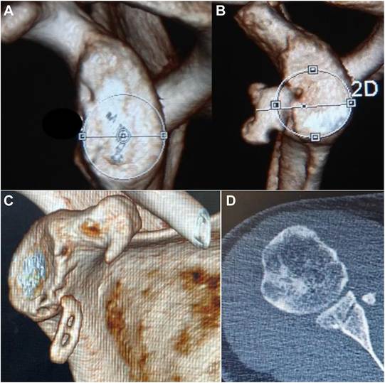
Figure 4 (A) Preoperative CT scan with glenoid bone loss. (B) Three-month follow-up CT after surgery demonstrating well-positioned and united coracoid graft. (C and D) Coracoid graft loosening with migration in a 3-month postoperative CT scan. CT, computed tomography.
| Table III Median values of preoperative and postoperative range of motion and scores using NPAR test, including the respective P values determined by Wilcoxon signed-ranked test. | imaging artifacts, reducing the chances of implant prominence, and reducing the risks of implant breakage being symptomatic. Cerclage FiberTape fixation of the coracoid graft was the newest innovation that aimed at reducing implant-related complications |
| PreOp FF A PostOp FF A PreOp ABD A PostOpABD A PreOp ER1 PostOp ER1 PreOp ER2 PostOp ER2 PreOp IR2 PostOp IR2 PreOp ASES PostOp ASES PreOp Walch Duplay PostOp Walch Duplay PreOp Rowe PostOp Rowe PreOp VAS PostOp VAS | 180.0 180.0 160.0 170.0 45.0 70.0 30.0 70.0 55.0 75.0 48.0 95.0 0.0 80.0 15.0 90.0 4.0 1.0 | 1.000 .180 .109 .102 .109 .109 .102 .109 .109 | 180.0 180.0 180.0 180.0 45.0 70.0 45.0 70.0 60.0 80.0 53.0 95.0 20.0 90.0 40.0 95.0 4.0 1.0 | .180 .157 .041 .034 .027 .018 .018 .018 .018 | analyzing the fixation strength of the cerclage FiberTape and follow-up studies following the cerclage FiberTape Latarjet are not available in the literature at present. The authors of the current study of cerclage FiberTape fixation assumed that the initial results following the procedure were not favorable due to increased rates of coracoid graft nonunion with migration. Thus, the hypothesis of the authors that cerclage FiberTape fixation could provide sturdy fixation of the coracoid graft was rejected. However, despite the high rates of graft loosening, none of the patients experienced any further episodes of redislocation until the date of follow-up. The authors consider that the remplissage procedure and capsulolabral tissue reattachment done along with the Latarjet helped in maintaining the stability of the shoulder. Long-term results of the Latarjet procedure with metal screws reported graft nonunion rates of around 9.1%.10 Hovelius et al reported 83% union rates with 14% graft migration in their long-term follow-up in open Latarjet with metal screws.14 Boileau et al in their follow-up study of Latarjet using cortical button reported 95% graft union rates with one case of graft migration3 and nonunion rates of 20% in another study by the same author.4 Graft migration was |
while also providing good graft union rates and functional outcomes following the procedure.12 The biomechanical studies calculated as a gap of 0.5 cm or more between the glenoid bed and
952
A.V. Nair, J.J. Panakkal, P.K. Mohan et al.
the coracoid graft.14 Our study using cerclage FiberTape for graft fixation demonstrated graft loosening and migration gaps in three of the total ten patients (30%), which were higher and more significant.
One of the salient findings that stood out were the good to excellent functional outcomes, irrespective of the status of coracoid graft union. Despite the small sample, the procedure employed in our study yielded functional outcomes that are at par with published literature on arthroscopic Latarjet using suspensory type of fixation.9 Apart from one patient who had a postoperative external rotation that was 5 degrees less than the preoperative value, all patients either regained or improved their external rotation. However, the mean postoperative ER1 and ER2 of the affected shoulder were less than those of the opposite shoulder, a deficit attributed to the remplissage,16 as well as the Latarjet procedure,4 which was not specifically complained of by any of the patients.
The authors reported that the chances of implant cut-out were negligible as the integrity of the drill holes in the coracoid graft was maintained in follow-up CT scans. The authors suspected several possibilities for early graft loosening. First, securing the FiberTape fixation in the posterior aspect of the glenoid was a blind procedure. The soft tissue interposition between the glenoid and the FiberTape may lead to early loosening. The authors suspect that fixation of the coracoid graft with cerclage FiberTape was not sturdy enough to withstand the aggressive fast-track rehabilitation protocol usually employed for their patients who have undergone Latarjet with screw fixation, probably being another factor accounting for high rate of graft loosening.
There were several limitations to this study. First, this was a single-center study with a limited number of patients operated on by a fellowship-trained shoulder surgeon. Second, this was a shortterm follow-up study. Long-term follow-ups to analyze graft remodeling and osteolysis are warranted. The authors concede that an additional CT scan in the immediate postoperative period would have enabled the differentiation between loosely apposed coracoid graft intraoperatively from postoperative graft loosening as well as distinguishing intraoperative malreduction from postoperative loss of reduction. More studies with longer-term follow-ups and a more significant number of patients were needed to decide on the future use of the cerclage FiberTape.
Conclusion
Our study demonstrated that arthroscopic Latarjet using cerclage FiberTape fixation combined with remplissage and capsulolabral repair resulted in a high rate of graft loosening and migration (30%). Nonetheless, patients in whom the coracoid graft had united, as well as those in whom it had not, all had good to excellent functional and clinical outcomes, no complications, and did not require any revision surgery.
Disclaimers:
Funding: No funding was disclosed by the authors.
Conflicts of interest: The authors, their immediate families, and any research foundation with which they are affiliated have not received any financial payments or other benefits from any commercial entity related to the subject of this article.
Supplementary Data
Supplementary data to this article can be found online at https://doi.org/10.1016/j.jseint.2024.03.020.
JSES International 8 (2024) 946e953 References
- Athwal GS, Meislin R, Getz C, Weinstein D, Favorito P. Short-term complications of the arthroscopic Latarjet procedure: a North American experience. Arthroscopy 2016;32:1965-70. https://doi.org/10.1016/j.arthro.2016.02.022.
- Balestro JC, Young A, Maccioni C, Walch G. Graft osteolysis and recurrentinstability after the Latarjet procedure performed with bioabsorbable screw fixation. J Shoulder Elbow Surg 2015;24:711-8. https://doi.org/10.1016/
- Boileau P, Saliken D, Gendre P, Seeto BL, d’Ollonne T, Gonzalez JF, et al. Arthroscopic Latarjet: suture-button fixation is a safe and reliable alternative to screw fixation. Arthroscopy 2019;35:1050-61. https://doi.org/10.1016/ j.arthro.2018.11.012.
- Boileau P, Thelu C E, Mercier N, Ohl X, Houghton-Clemmey R, Carles M, et al. Arthroscopic Bristow-Latarjet combined with bankart repair restores shoulder stability in patients with glenoid bone loss. Clin Orthop 2014;472:2413-24. https://doi.org/10.1007/s11999-014-3691-x.
- Chaudhary D, Goyal A, Joshi D, Jain V, Mohindra M, Mehta N. Clinical andradiological outcome after mini-open Latarjet technique with fixation of coracoid with Arthrex wedge mini-plate. J Clin Orthop Trauma 2016;7:23-9. https://doi.org/10.1016/j.jcot.2015.09.002.
- Dumont GD, Fogerty S, Rosso C, Lafosse L. The arthroscopic Latarjet procedurefor anterior shoulder instability: 5-year minimum follow-up. Am J Sports Med 2014;42:2560-6. https://doi.org/10.1177/0363546514544682.
- Frank RM, Roth M, Wijdicks CA, Fischer N, Costantini A, Di Giacomo G, et al.Biomechanical analysis of plate fixation compared with various screw configurations for use in the Latarjet procedure. Orthop J Sports Med 2020;8: 2325967120931399. https://doi.org/10.1177/2325967120931399.
- Giacomo GD, Costantini A, de Gasperis N, De Vita A, Lin BKH, Francone M, et al.Coracoid bone graft osteolysis after Latarjet procedure: a comparison study between two screws standard technique vs mini-plate fixation. Int J Shoulder Surg 2013;7:1-6. https://doi.org/10.4103/0973-6042.109877.
- Girard M, Dalmas Y, Martinel V, Mansat P, Bonnevialle N. Arthroscopic Latarjetwith cortical buttons versus open Latarjet with screws: a short-term comparative study. Am J Sports Med 2022;50:3326-32. https://doi.org/10.1177/ 03635465221120076.
- Griesser MJ, Harris JD, McCoy BW, Hussain WM, Jones MH, Bishop JY, et al.Complications and re-operations after Bristow-Latarjet shoulder stabilization: a systematic review. J Shoulder Elbow Surg 2013;22:286-92. https://doi.org/ 10.1016/j.jse.2012.09.009.
- Gupta A, Delaney R, Petkin K, Lafosse L. Complications of the Latarjet procedure. Curr Rev Musculoskelet Med 2015;8:59-66. https://doi.org/10.1007/ s12178-015-9258-y.
- Hachem AI, Costa D’O G, Rondanelli SR, Rius X, Barco R. Latarjet cerclage: the metal-free fixation. Arthrosc Tech 2020;9:e1397-408. https://doi.org/10.1016/ j.eats.2020.05.024.
- Hardy A, Sabatier V, Schoch B, Vigan M, Werthel JD. Latarjet with corticalbutton fixation is associated with an increase of the risk of recurrent dislocation compared to screw fixation. Knee Surg Sports Traumatol Arthrosc 2020;28:2354-60. https://doi.org/10.1007/s00167-019-05815-6.
- Hovelius L, Sandstrom B, Olofsson A, Svensson O, Rahme H. The effect of€ capsular repair, bone block healing, and position on the results of the BristowLatarjet procedure (study III): long-term follow-up in 319 shoulders. J Shoulder Elbow Surg 2012;21:647-60. https://doi.org/10.1016/j.jse.2011.03.020.
- Itoi E. ‘On-track’ and ‘off-track’ shoulder lesions. EFORT Open Rev 2017;2:343-
51. https://doi.org/10.1302/2058-5241.2.170007.
- Ko SH, Cha JR, Lee CC, Hwang IY, Choe CG, Kim MS. The influence of arthroscopic remplissage for engaging Hill-Sachs lesions combined with Bankart repair on redislocation and shoulder function compared with Bankart repair alone. Clin Orthop Surg 2016;8:428-36. https://doi.org/10.4055/ cios.2016.8.4.428.
- Ladermann A, Denard PJ, Burkhart SS. Injury of the suprascapular nerve during€ Latarjet procedure: an anatomic study. Arthroscopy 2012;28:316-21. https:// doi.org/10.1016/j.arthro.2011.08.307.
- Lafosse L, Boyle S. Arthroscopic Latarjet procedure. J Shoulder Elbow Surg2010;19(2 Suppl):2-12. https://doi.org/10.1016/j.jse.2009.12.010.
- Mizuno N, Denard PJ, Raiss P, Melis B, Walch G. Long-term results of theLatarjet procedure for anterior instability of the shoulder. J Shoulder Elbow Surg 2014;23:1691-9. https://doi.org/10.1016/j.jse.2014.02.015.
- Provencher MT, Bhatia S, Ghodadra NS, Grumet RC, Bach BR, Dewing CB, et al.Recurrent shoulder instability: current concepts for evaluation and management of glenoid bone loss. J Bone Joint Surg Am 2010;92(Suppl 2):133-51. https://doi.org/10.2106/JBJS.J.00906.
- Skupinski J, Piechota MZ, Wawrzynek W, Maczuch J, Babi nska A. The Bony Bankart lesion: how to measure the glenoid bone loss. Pol J Radiol 2017;82:5863. https://doi.org/10.12659/PJR.898566.
- Valenti P, Maroun C, Wagner E, Werthel JD. Arthroscopic Latarjet procedurecombined with Bankart repair: a technique using 2 cortical buttons and specific glenoid and coracoid guides. Arthrosc Tech 2018;7:e313-20. https://doi.org/ 10.1016/j.eats.2017.09.009.
- Walch G, Boileau P. Latarjet-Bristow procedure for recurrent anterior instability. Tech Shoulder Elb Surg 2000;1:256-61.