Contributors
Ayyappan V. Nair, M.B.B.S., D.N.B., Jetty Tejaswari, M.B.B.S., M.S., Prince Shanavas Khan, M.S., Kiran Veerendra, M.S., M.R.C.S., Aravind Rajan, M.B.B.S., M.S., D.N.B., Shakir Rashid, M.B.B.S., M.S., and
M. Priyamvada, B.Tech, M.Tech
Summary
Arthroscopy Techniques
Link: https://doi.org/10.1016/j.eats.2025.103961
Glenoid articular cartilage lesions are a significant cause of persistent shoulder pain, particularly following trauma or previous surgical interventions, such as failed Bankart repairs. Current surgical approaches for addressing cartilage damage in the glenohumeral joint include microfracture, osteochondral autograft transfer, osteochondral allograft transplantation, and autologous chondrocyte implantation. However, the use of bone marrow aspiration concentrate (BMAC), which is rich in mesenchymal stem cells, has emerged as a promising minimally invasive singlestep procedure that promotes cartilage regeneration. This study presents an arthroscopic technique for addressing cartilage defects in the glenoid using BMAC after failed Bankart repair in a patient. Although the evidence base for BMAC in glenoid defects is limited, early clinical results suggest effective cartilage healing, few complications, and satisfactory patient outcomes. This technique appears to be safe and offers the benefit of being a single-step process that utilizes autologous bone marrow mesenchymal cells to facilitate cartilage regeneration. Further research is needed to establish the long-term efficacy of this approach for treating glenoid cartilage lesions.
| G |
lenoid articular cartilage lesions are a recognized significant functional limitation and may not always be cause of persistent shoulder pain, particularly readily apparent on magnetic resonance imaging
| following trauma or previous surgical interventions. In 1993, Neviaser was the first to identify the glenolabral articular disruption lesion, describing it as a traumatic injury that leads to a tear in the anterior inferior labrum and a loss of articular cartilage, which results in pain in the front of the shoulder.1 Although more prevalent in load-bearing joints, such as the knee and ankle, glenoid chondral defects can be a source of | (MRI). Their association with conditions such as shoulder instability, repetitive microtrauma, or previous failed surgeries, such as Bankart repair, has been well established. 2-4 Overhead athletes and patients with rotator cuff pathology frequently exhibit glenoid cartilage lesions, with studies reporting incidences ranging from 13% to 57% depending on the associated pathology and patient population. 5 Cartilage defects were commonly found in 38% of |
patients with anterior glenohumeral instability.6 The etiology of these lesions includes trauma, which can be a single event or repetitive microtrauma, postarthroscopic surgery, infections, osteochondritis dissecans, and an association with instability and rotator cuff tears.7
The literature on treatment options and outcomes for full-thickness glenoid articular lesions is notably sparse. Current surgical approaches for addressing cartilage damage in the glenohumeral joint include microfracture, osteochondral autograft transfer,8 osteochondral allograft transplantation (OCA), and autologous chondrocyte implantation.9 These techniques, initially
Arthroscopy Techniques, Vol 14, No 12 (December), 2025: 103961 e1
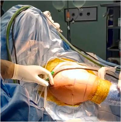
Fig 1. Patient placed in the beach-chair position.
developed for managing chondral lesions in weightbearing joints, have been adapted for shoulder use.
Role of Bone Marrow Aspiration Concentrate
Bone marrow aspiration concentrate (BMAC) is rich in mesenchymal stem cells (MSCs) capable of both direct chondrogenic differentiation and paracrine signaling, which stimulate cartilage repair. Because MSCs comprise only 0.001% to 0.02% of all nucleated cells in unprocessed bone marrow aspirate, commercially available systems use density-gradient centrifugation to enhance their concentration by up to 5-fold, resulting in BMAC, which is then mixed with thrombin solution. This mixture is drawn into a syringe, and another syringe with fibrin sealant is prepared. Both syringes are attached to a 20-gauge application needle using a Y-shaped connector), allowing BMAC and fibrin glue to be delivered simultaneously during application from a scaffold, which helps to retain BMAC within the defect. The use of autologous BMAC offers several advantages, including the following:
- Minimally invasive, single-step procedure
- No donor site morbidity
- Use of the patient’s own MSCs, reducing immunologic risks
- Promotes cartilage regeneration via both differentiation and paracrine mechanisms
Although the evidence base for BMAC in glenoid defects is limited, early clinical results suggest effective cartilage healing, few complications, and satisfactory patient outcomes.
Recognizing the link between focal chondral defects and shoulder instability, we were motivated to document our technique because of the lack of existing literature on an arthroscopic approach for repairing chondral damage of the glenoid during the arthroscopic Latarjet procedure.
Surgical Technique
A video presenting arthroscopic method for addressing glenoid cartilage damage (Video 1).
We present an arthroscopic method for addressing glenoid cartilage damage following unsuccessful Bankart repair. All participants provided informed consent. Preoperative imaging includes standard radiography and MRI to assess the size and position of the cartilage defect and identify any additional related conditions.
Patient Setup and Positioning
The patient is placed in a beach-chair position (Fig 1) under general anesthesia and an interscalene block in the beach-chair position. A traction force of 5 kg is applied to the forearm, maintaining the arm at a 60◦
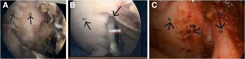
Fig 2. Arthroscopic visualization from the suprabicipital portal in the right shoulder with a patient in the beach-chair position shows glenoid catilage defect (A), defect meticulously debrided with a curette (B), and microfracturing of the defect done to ensure BMAC retention and maximize cell activity for regeneration (C). (BMAC, bone marrow aspirate concentrate; G, glenoid; GC, glenoid cartilage; MF, microfractured defect area.)
TREATING GLENOID CARTILAGE DEFECTS WITH BMAC e3
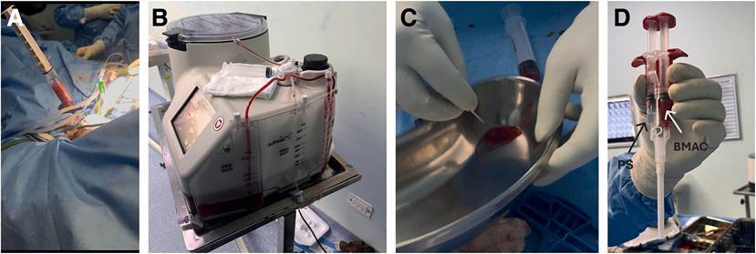
Fig 3. (A) Bone marrow aspiration from 3 finger breadths above the anterosuperior iliac spine. (B) Total of 60 mL bone marrow aspirated and processed in the Arthrex system. (C) Bone marrow aspirate is mixed with thrombin solution. (D) The mixture is drawn into the syringe. Another syringe with sealant protein is prepared, and both syringes are attached to a 20-gauge application needle using a Y-shaped connector.
elevation, with 10 ◦ of shoulder abduction and in a neutral rotation. The right shoulder is prepared using 7.5% povidone-iodine and covered with sterile drapes. Similarly, the area from the right iliac crest to the inguinal region is prepared for bone marrow aspiration.
Diagnostic arthroscopy is initiated using the posterior portal. An arthroscopic Latarjet procedure is performed using 2 cortical buttons (Smith & Nephew. Andover, MA), one placed anteriorly and the other posteriorly.
Attention is then directed toward addressing the cartilage defect located anteriorly in the glenoid (Fig 2A). The bed of the defect, identified as an Outerbridge grade 4 cartilage defect, is debrided with a curette (Fig 2B). The cartilage defect is microfractured (Fig 2C), and bone marrow is aspirated percutaneously from the right iliac crest by inserting the trocar of the bone marrow aspiration kit (Arthrex. Naples, FL) approximately 3 finger breadths proximal to the anterior superior iliac spine
Table 1. Different Steps of the Operation
- Patient placed in a beach-chair position under general anesthesia and an interscalene block
- Diagnostic arthroscopy performed
- Arthroscopic Latarjet and remplissage performed
- Anterior glenoid defect visualized
- Defect bed debrided with curette and microfracturing of defect done
- Bone marrow aspiration: percutaneous aspiration from the
right iliac crest, trocar inserted 3 finger breadths above the anterosuperior iliac spine
- 60 mL bone marrow aspirate processed using the Arthrex Angel Concentrated Platelet Rich Plasma System
- Defect area dried, joint maintained in a dry field with CO2 insufflation (15 mm Hg, 2 L/min)
- BMAC concentrate mixed with human thrombin
- BMAC and fibrin glue delivered simultaneously during application to form a scaffold
BMAC, bone marrow aspirate concentrate.
(Fig 3A). A total of 60 mL of bone marrow aspirate is collected and processed using an Angel Concentrated Platelet Rich Plasma System (Arthrex) (Fig 3B). The prepared cartilage defect area is dried and maintained in a dry field to ensure adequate joint distension. Carbon dioxide insufflation is performed at a pressure of 15 mm Hg and a flow rate of 2 L/min, a method that has been previously shown to be safe in knee arthroscopy. The final BMAC concentrate is mixed with human thrombin (Baxter, Vienna, Austria) (Fig 3C) and applied along with sealer protein concentrate (Baxter), which together form fibrin glue to cover the cartilage defect (Fig 3D). No additional washing is performed, allowing the scaffold with the marrow concentrate to form a compact plug (Fig 4).
Rehabilitation
The shoulder is initially immobilized postoperatively.
Early motion of the elbow and hand begins on day 1; passive shoulder exercises commence on day 10. Full
passive range of motion is achieved by 6 weeks, with progression to strengthening thereafter. Return to daily activity is permitted at 6 weeks, with a gradual resumption of sports activities after 6 months. The addition of cartilage repair does not require deviation from standard rehabilitation following the Latarjet and remplissage procedures.
Discussion
Chondral lesions of the glenoid are frequently missed during preoperative imaging and may present as persistent pain following stabilization. Accurate diagnosis using advanced imaging techniques, such as MRI, is crucial for selecting the most appropriate treatment approach, including surgical interventions.
Optimal management aims to restore the glenoid articular surface and geometry, minimize the exposed articular defect, re-establish stability, and preserve long-term function.10 The choice of joint-preserving treatment for younger and more active individuals remains a topic of discussion.
Among the available techniques, BMAC stands out as a safe, autologous, and single-step intervention that promotes hyaline-like cartilage repair in small case series and animal models. Table 2 summarizes the therapeutic approaches for glenohumeral joint articular cartilage lesions in young individuals.
Millet et al. 11 examined 25 shoulders that received microfracture treatment for articular cartilage injuries. They observed a notable improvement in 81% of the shoulders (25/31), with the most significant progress seen in smaller lesions, whereas bipolar lesions had poorer outcomes. A drawback of this method is the formation of fibrocartilage instead of hyaline cartilage. Fibrocartilage is less advantageous than hyaline cartilage in terms of joint surface mechanics.
Table 2. Therapeutic Approaches for Glenohumeral Joint Articular Cartilage Lesions in Young Individuals
| Category | Procedure | Indications | Advantages | Limitations |
| Arthroscopic surgery | Debridement and lavage | Small, superficial, or frayed lesions | Minimally invasive, short recovery | Not restorative; symptom control only |
| Microfracture | Small (<2-4 cm 2 ), full-thickness unipolar defects | Stimulates fibrocartilage repair | Fibrocartilage inferior to hyaline; unpredictable durability | |
| Cartilage restoration | Osteochondral autograft transfer | Small unipolar lesions in young patients | Transplants native hyaline cartilage | Donor site morbidity, technically challenging |
| Osteochondral allograft transplantation | Larger unipolar/bipolar lesions; failed prior surgery | Replaces bone and cartilage; no donor site morbidity | Graft availability, risk of nonintegration | |
| Autologous chondrocyte implantation | Larger full-thickness lesions (mostly investigational in shoulder) | Regeneration with hyaline-like tissue | Two-stage procedure, limited shoulder data | |
| Biologic resurfacing | Fascia lata autograft, dermal allograft, scaffold | Bipolar lesions with preserved joint space (young patients) | Joint-preserving, delays arthroplasty | Variable outcomes, longterm durability uncertain |
TREATING GLENOID CARTILAGE DEFECTS WITH BMAC e5
Table 3. Advantages and Disadvantages
Advantages
One-step procedure
More than cost-effective than ACI
Fully arthroscopic procedure
Promotes hyaline-like cartilage repair
Disadvantages
No long-term follow-up
Limited high-level evidence
ACI, autologous chondrocyte implantation.
In their systematic review, Prigmore et al.12 explored the application of OCA transplantation for addressing traumatic impaction fractures, recurrent instability, and degenerative joint disease in the shoulder. The review encompassed 17 studies with a total of 83 shoulders, indicating an overall success rate of 82% and generally positive outcomes, especially for isolated osteochondral injuries. These results imply that OCA is a promising biological treatment option for complex shoulder conditions, although the evidence is largely derived from case reports and series. The main drawbacks are the availability of grafts and the risk of integration.
According to Riff et al.,13 OCA is a feasible option for young individuals with isolated chondral damage in the humerus, although the presence of bipolar disease in the shoulder leads to higher failure rates and poorer outcomes.
Pham et al. 14 documented a case involving a 14-yearold adolescent with osteochondritis dissecans in the humeral head, which was successfully treated using an arthroscopic osteochondral autograft transfer from the knee, resulting in a satisfactory outcome. The main drawback of this treatment is donor site morbidity.
Buchmann et al.15 documented the treatment of 3 full-thickness cartilage defects in the humerus (each measuring 6.0 cm2 ) and 1 full-thickness cartilage defect in the glenoid (2.0 cm2 ) using autologous chondrocyte implantation. Postsurgery, the average visual analog scale score was 0.3 of 10, the mean Constant score was 83.3 ± 9.9, and the average American Shoulder and Elbow Surgeons index was 95.3 ± 8.1, indicating satisfactory shoulder function. MRI results showed adequate coverage of the defects, with evidence of fibrocartilaginous repair tissue.
Table 4. Pears and Pitfalls of Our Technique
Pearls
Single-step repair
Autologous source
Density gradient centrifugation: increases yield of mesenchymal stem cells, boosting chondrogenic potential of BMAC
Meticulous defect preparation ensures BMAC retention and maximizes cell activity for regeneration
Pitfalls
Technically challenging
Fluid dilution and washout Limited long-term data
BMAC, bone marrow aspirate concentrate.
Fortier et al. 16 conducted a comparative study on the
outcomes of treatment using BMAC versus micro-
fracture alone in an equine model of osteoarthritis. The findings indicated that BMAC resulted in significantly superior outcomes both macroscopically and histologically after 3 months. Additionally, the combination of BMAC with microfracturing led to an increased production of type 2 collagen and glycosaminoglycans in the repaired tissue.
In a recent case report, Nair et al. 17 described the efficacy of arthroscopic BMAC application in treating cartilage defects of the humeral head and validated its technical feasibility and satisfactory clinicoradiological outcomes when used in conjunction with Latarjet, capsulolabral repair, and remplissage arthroscopically.
In the knee, BMAC has shown effectiveness in treating chondral lesions, with reports indicating an improvement in the visual analog score from 5.4 to 0.5 and the International Knee Documentation Committee Subjective Knee Form score from 39.2 to 82.2. Notably, follow-up MRI results showed excellent defect filling in 81% of patients younger than 45 years and 70% of those older than 45 years. 18 Similarly, in cases of talar chondral lesions, MRI indicated that 77.3% of defects were completely filled, and there was a significant enhancement in the Foot and Ankle Outcome Score. 19
To date, research on the application of BMAC for glenoid cartilage lesions has been limited. Nevertheless, the few available studies have shown encouraging outcomes, indicating effective cartilage repair and overall improvement without an increase in adverse effects compared with other cartilage repair methods.
This technique appears to be safe, offering the benefit of being a single-step process that utilizes autologous bone marrow mesenchymal cells to aid cartilage regeneration (Tables 3, 4).
Disclosures
All authors (A.V.N., J.T., P.S.K., K.V., A.R., S.R., P.M.) declare that they have no known competing financial interests or personal relationships that could have appeared to influence the work reported in this paper.
Acknowledgments
During the preparation of this work, the authors used Wondershare Filmora 14 version in order to edit and narrate the video.
References
- Neviaser TJ. The GLAD lesion: Another cause of anterior shoulder pain. Arthroscopy 1993;9:22-23.
- Løken S, Granan LP, Sivertsen EA, Årøen A. Epidemiology of cartilage injuries. In: Sports Injuries. Berlin, Heidelberg: Springer, 2013;1-12.
- Carroll KW, Helms CA, Speer KP. Focal articular cartilage lesions of the superior humeral head. Am J Roentgenol 2001;176:393-397.
- Guntern DV, Pfirrmann CWA, Schmid MR, et al. Articular cartilage lesions of the glenohumeral joint: Diagnostic effectiveness of MR arthrography and prevalence in patients with subacromial impingement syndrome. Radiology 2003;226:165-170.
- Ruckstuhl H, de Bruin ED, Stussi E, Vanwanseele B. Posttraumatic glenohumeral cartilage lesions: A systematic review. BMC Musculoskelet Disord 2008;9:107.
- Kawakami J, Yamamoto N, Itoi E, Henninger H,
Tashjian R, Chalmers PN. Morphology of glenoid cartilage defects in anteroinferior glenohumeral instability. Orthop J Sports Med 2022;10:23259671221086615.
- Dagher D, Selznick A, Prada C, Al Shehab Y, Leroux T, Khan M. Surgical treatment options for articular cartilage defects of the glenohumeral joint: A systematic review. Shoulder Elbow 2023;15:580-592.
- Mitchell JJ, Vap AR, Sanchez G, et al. Concomitant reverse Hill-Sachs lesion and posterior humeral avulsion of the glenohumeral ligament: Treatment with fresh talus osteochondral allograft and arthroscopic posterior humeral avulsion of the glenohumeral ligament and labrum repair. Arthrosc Tech 2017;6:e987-e995.
- Flatow EL, Altchek DW, Gartsman GM, et al. Commentary. Orthop Clin North Am 1997;28:277-294.
- Porcellini G, Cecere AB, Giorgini A, Micheloni GM, Tarallo L. The GLAD lesion: Are the definition, diagnosis and treatment up to date? A systematic review. Acta Biomed Mattioli 1885 2020;91:1-9.
- Millett PJ, Huffard BH, Horan MP, Hawkins RJ, Steadman JR. Outcomes of full-thickness articular cartilage injuries of the shoulder treated with microfracture. Arthroscopy 2009;25:856-863.
- Prigmore B, Tabbaa S, Crawford DC. Osteochondral allograft transplantation in the shoulder: A systematic review of indications and outcomes. Cartilage 2024;15:219-228.
- Riff AJ, Yanke AB, Shin JJ, Romeo AA, Cole BJ. Midterm results of osteochondral allograft transplantation to the humeral head. J Shoulder Elbow Surg 2017;26:e207-e215.
- Pham TT, Kany J, Lakhal W, Sales de Gauzy J, Accadbled F. Arthroscopic osteochondral autograft transfer for juvenile osteochondritis dissecans of the humeral head. JBJS Case Connect 2017;7:e63.
- Buchmann S, Salzmann GM, Glanzmann MC, Wörtler K,
Vogt S, Imhoff AB. Early clinical and structural results after autologous chondrocyte transplantation at the glenohumeral joint. J Shoulder Elbow Surg 2012;21:1213-1221.
- Fortier LA, Potter HG, Rickey EJ, et al. Concentrated bone marrow aspirate improves full-thickness cartilage repair compared with microfracture in the equine model. J Bone Joint Surg Am 2010;92:1927-1937.
- Nair AV, Rambhojun M, Sreejith Thampy J, Khan PS. Arthroscopic technique for management of unusual cartilage defect in the shoulder with BMAC (bone marrow aspiration concentrate)―a case report. J Orthop Rep 2024;3:100311.
- Gobbi A, Scotti C, Karnatzikos G, Mudhigere A, Castro M, Peretti GM. One-step surgery with multipotent stem cells and hyaluronan-based scaffold for the treatment of fullthickness chondral defects of the knee in patients older than 45 years. Knee Surg Sports Traumatol Arthrosc 2017;25: 2494-2501.
- Hannon CP, Ross KA, Murawski CD, et al. Arthroscopic bone marrow stimulation and concentrated bone marrow aspirate for osteochondral lesions of the talus: A casecontrol study of functional and magnetic resonance observation of cartilage repair tissue outcomes. Arthroscopy 2016;32:339-347.