

Arun Kumar Chinraja, Azad Saita, Aebel Rajub, Jimmy Joseph Meleppuramb, Ajayakumar Thankappanb, Ayyappan V. Nairc, Prince Shanavas Khanb,*
- Journal of ISAKOS – 2024
LINK: https://www.jisakos.com/article/S2059-7754(24)00093-2/fulltext
This report describes a novel surgical approach for isolated anterior branch axillary nerve injury using an upper and middle trapezius transfer to the anterior deltoid via a subacromial route, facilitated by an autologous semitendinosus graft. A 55-year-old male presented with progressive deltoid wasting and weakness following a shoulder dislocation 18 months prior, with limited abduction (120°) and reduced deltoid power (grade 3). Imaging showed deltoid atrophy without rotator cuff injury, and electromyography confirmed selective anterior branch axillary nerve involvement. Surgical exploration revealed a non-stimulable anterior branch, leading to trapezius tendon harvest, augmentation with semitendinosus graft, and transfer under the acromion to the deltoid insertion. Postoperative rehabilitation included abduction splinting and gradual mobilization. At four years follow-up, the patient demonstrated significant improvement in shoulder function, with abduction increasing to 160° and deltoid power to grade 4+, enabling return to manual work. This technique offers advantages over traditional methods by avoiding osteotomy, reducing morbidity, and allowing earlier rehabilitation. It provides a valuable option for chronic, branch-specific axillary nerve injuries where nerve repair is not feasible, emphasizing restoration of muscle congruence for optimal functional recovery.
* Corresponding author. Tel.: þ919446503401.
E-mail address: drpskhan@gmail.com (P.S. Khan).
Received 7 December 2023; Received in revised form 16 April 2024; Accepted 8 May 2024
Available online 11 May 2024
2059-7754/© 2024 The Author(s). Published by Elsevier Inc. on behalf of International Society of Arthroscopy, Knee Surgery and Orthopedic Sports Medicine. This is an open access article under the CC BY-NC-ND license (http://creativecommons.org/licenses/by-nc-nd/4.0/).
INTRODUCTION
The vulnerability of the anterior branch of the axillary nerve near the surgical neck of the humerus is well-known but often under-addressed in literature. In response, an approach involving the transfer of the upper and middle trapezius to the anterior deltoid via a subacromial path, facilitated by an autologous semitendinosus graft, has been introduced. This technique, aimed at restoring anterolateral deltoid fiber congruence, offers a viable option for patients with isolated anterior branch axillary nerve paralysis, particularly in cases where conventional treatments may have limited effectiveness, including nerve grafting, shoulder arthrodesis, and described techniques involving osteotomy [1,2]. Despite chronicity, functional improvements have been observed, mitigating the need for more morbid interventions like humerus osteotomy or shoulder arthrodesis. This underscores the importance of tailored approaches to nerve injuries, emphasizing the restoration of muscle integrity for optimal recovery.
CASE REPORT
A 55-year-old male patient presented to our clinic with a 5-month history of progressive reduction in muscle mass and weakness in the left shoulder, particularly during abduction, accompanied by a gradual decline in muscle mass and alteration in shoulder contour. He recounted an incident 18 months prior when he experienced a shoulder dislocation while losing balance while unloading a heavy load. Seeking the assistance of a traditional bone setter, the dislocation was reduced, but subsequent months were marked by persistent pain, mitigated with selfadministered oral analgesics. He had no history of cerebrovascular accidents, recent traumatic events or other swellings.
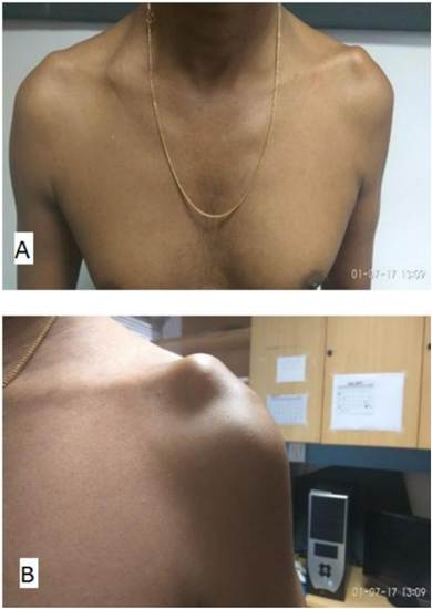
The general physical examination was within normal limits. During the left shoulder examination, we observed reduced muscle mass in the anterior deltoid region (Fig. 1A, B). The patient had limited active shoulder abduction, reaching 120, and forward elevation limited to 70 with grade 3 deltoid power and grade 5 trapezius power (MRC grading). Importantly, no signs of shoulder impingement or rotator cuff injury were found. The latissimus dorsi and teres minor muscles showed normal function).
Imaging
Plain radiographs indicated a dislocated shoulder with a slight inferior sag (Fig. 2A). Magnetic resonance imaging scan revealed deltoid muscle fatty infiltration and atrophy, while cuff muscles and the brachial plexus appeared normal, no mass lesion was found (Fig. 2B). Nerve conduction studies indicated left axillary axonal neuropathy. An axillary artery angiogram ruled out quadrangular space syndrome [3].
Electrophysiological analysis
Electromyography (EMG) confirmed denervation in the anterior, lateral deltoid area, while posterior deltoid function remained unaffected. The diagnosis was isolated anterior branch axillary nerve injury.
Surgical technique
Given the duration of the injury, a surgical plan was devised, involving the exploration and neurolysis of the axillary nerve. Additionally, decompression of the quadrilateral space and intraoperative
Fig. 1. A, B: Deltoid muscle wasting over the left deltoid region.
stimulation of the nerve branches were planned. If the anterior branch exhibited stimulability, the plan was to conclude with neurolysis; otherwise, an upper trapezius transfer was intended.
The patient was positioned in right lateral decubitus, and the left upper limb and leg were prepared and draped for surgery [4]. An incision, resembling a mirrored lazy “S,” commenced above the acromion, curving along the posterior border of the deltoid, and extending below its insertion (Fig. 3). Thick skin flaps were raised on both sides, with a cautious dissection along the posterior skin flap to identify the cutaneous branch of the axillary nerve (Fig. 4A). Subsequently, tracing led to the quadrangular space for identification of the nerve to the teres minor, along with the posterior and anterior branches. While the posterior branch displayed stimulability, the anterior branch did not. Consequently, the decision was made to proceed with the harvest of the upper trapezius transfer.
We then traced the trapezius muscle insertion on the acromion and detached the trapezius tendon along with the periosteum (Fig. 4B). The semitendinosus autograft was harvested, and the procedure involved executing it through a small posterior incision along its course using an open tendon stripper. The donor muscle tendon was pulvertafted using the semitendinosus graft to attain the necessary length. Subsequently, it was passed beneath the acromion along the anterior portion of the deltoid to reach its insertion point, securing it in place with a biotenodesis screw while maintaining the upper arm abducted to approximately 45 (Figs. 5 and 6).
Postoperative management and rehabilitation
In the postoperative phase, the patient was given an abduction splint set at 30 for 8 weeks. Passive shoulder abduction exercises commenced after 2 weeks, and active shoulder abduction was allowed after 6 weeks postoperatively. Over four years of follow-up, the patient demonstrated significant improvement. His abduction power increased from grade 3 preoperatively to grade 4þ in the scapular plane (MRC grading), and the degree of abduction improved from 120 preoperatively to 160 postoperatively (Fig. 7).
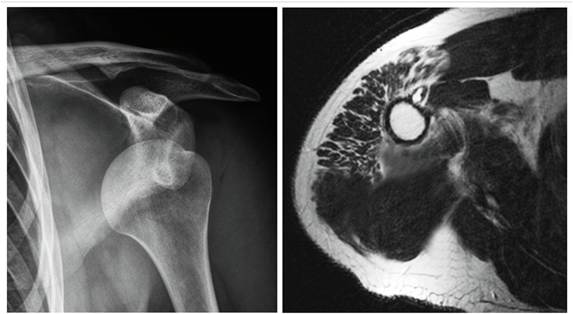
Fig. 2. Figure A (left) radiograph and B (right) MRI image of the patient showing atrophy of the anterior and middle deltoid muscle. MRI ¼ magnetic resonance imaging.
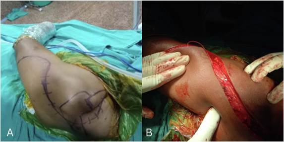
Fig. 3. A, B: Mirrored lazy “S” made starting above the acromion and extending down to below the deltoid insertion.
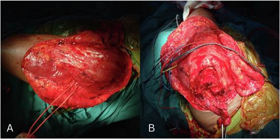
Fig. 4. A, B: A) Identification of the cutaneous sensory branch of the axillary nerve, tracing it to the quadrangular space. B) Trapezius muscle insertion on the acromion and detached the trapezius tendon along with the periosteum.
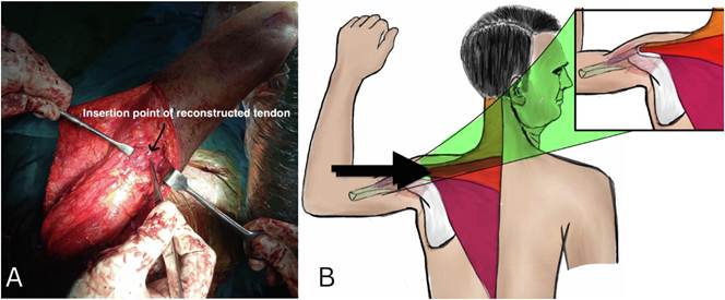
Fig. 5. A, B: The trapezius tendon and a contralateral semitendinosus muscle graft were harvested, pulvertafted, and secured near the deltoid muscle insertion with a
bioabsorbable screw.
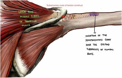
Fig. 6. Final construct of the semitendinosus muscle graft routed under the acromion process to the deltoid insertion.
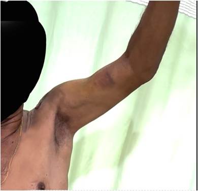
Fig. 7. Postoperative image showing the range of abduction for the patient.
Table 1
Etiology and description of axillary nerve injuries.
| Etiology | Description |
| Traumatic glenohumeral events | Result from shoulder trauma, sometimes involving humeral head subluxation.Nerve compression, stretching, or bruising under the deltoid muscle is often linked to accidents (e.g. car crashes) and contact sports (e.g. skiing, football, rugby). |
| Repetitive microtraumatic stress | – Occurs due to recurrent minor injuries that accumulate over time. Common among athletes who repeatedly perform overhead throwing motions, such as baseball pitchers. |
| Neuritis | – Inflammatory conditions affecting the brachial plexus, including the axillary nerve, can result in injury. |
| Quadrilateral space impingement | – Axillary nerve compression within the quadrilateral space of the shoulder can lead to injury. |
| Blunt force trauma | – Direct impact or trauma to the shoulder region can cause damage to the axillary nerve. |
| Neural entrapment due to mass lesions | – Space-occupying lesions like tumors or cysts can exert pressure on or entrap the axillary nerve, potentially causing injury. |
DISCUSSION
Isolated axillary nerve injuries are sparse and are frequently linked to brachial plexus injuries. The typical causes are listed (Table 1). The use of axillary nerve neurolysis and an innovative upper trapezius to anterior deltoid transfer via a subacromial path posterior to the clavicle, facilitated by an autologous semitendinosus graft, resulted in significant improvement with 140 degrees of forward flexion and Grade 4 power (MRC grading) at the 4-year follow-up.
Interestingly, the clinical symptoms of branch-specific axillary nerve injury, such as limited range of motion and pain, can make it difficult to detect during the initial evaluation. This is because the weakness in shoulder abduction is often compensated for by the supraspinatus muscle’s inherent ability to perform abduction [5].
EMG findings indicated reduced amplitude and denervation in the anterior along with the lateral deltoid, strongly suggesting an involvement of the axillary nerve. Surprisingly, the posterior deltoid, infraspinatus, and teres minor muscles functioned normally. This contrast indicated selective impairment within the axillary nerve’s branches. Furthermore, it became evident that the anterior branch of the axillary nerve was primarily affected, while the posterior branch remained functional.
Commonly practiced approaches for the brachial plexus and its branch injuries are described. Neurolysis removes scar tissue from the axillary nerve, which is best for entrapment-related injuries. Neurorrhaphy is for acute cases with minimal scarring, and nerve grafting is considered when repair isn’t possible. For extensive scarring, nerve grafting, or neurotization with options like a triceps-to-axillary transfer can be effective. Given the patient’s difficulty in daily activities like combing hair and lifting weights due to weak shoulder abduction, a treatment plan was procured. Nerve grafting wasn’t an option due to the prolonged injury duration (over 18 months) [6].
In the initial stages following an injury, nerve grafting and other repair techniques, in conjunction with physiotherapy, are typically the primary modalities of treatment. In chronic nerve injuries with limited regenerative potential, options like muscle transfers, humerus osteotomy, or shoulder arthrodesis are considered. However, our patient exhibited normal flexion, abduction, and a passive range of motion up to 150, along with active abduction of 120. Therefore, humerus osteotomy and shoulder arthrodesis should only be considered last-resort options to be opted for.
In our case, electromyography findings indicated an isolated axillary nerve injury, while the suprascapular nerve, long thoracic nerve, and thoracodorsal nerve remained intact. The shoulder’s external rotators, such as the infraspinatus muscle, contributed to external rotation at 0 degrees of abduction, while the teres minor facilitated external rotation at 90. Despite comprehensive reconstruction efforts, published cases have shown an improvement in abduction of approximately 40% [7].
For near-full abduction recovery in isolated branch-specific axillary nerve palsy, restoring anterolateral deltoid fiber congruence is the key. In our patient, pectoralis major with coracobrachialis for flexion and posterior deltoid fibers with latissimus dorsi for extension were intact, indicating an isolated anterior branch injury.
This operative technique involves passing the trapezius tendon under the acromion process and pulvertafting it to the semitendinosus tendon at the deltoid insertion on the humerus offers distinct advantages over traditional methods such as the Meyer technique, Bateman modification of Meyer [8], and Saha’s technique [9]. Techniques involving acromion osteotomy have higher chances of injuring accessory spinal nerves and nonunion of the acromion following the repair [10]. Advantages of this technique include simplicity, shorter surgery duration, quicker postoperative rehabilitation, avoidance of unnecessary osteotomies, and reduced morbidity at the osteotomy site [4].The patient expressed profound satisfaction with the care received for their delayed anterior branch axillary nerve injury. The innovative surgical approach restored the individual’s shoulder function and surpassed expectations, enabling a return to manual laborer with enhanced strength and mobility, ultimately contributing to a significant improvement in overall quality of life.
Declaration of competing interest
The authors declare that no financial interests/personal relationships exist to be considered as potential competing interests.
Acknowledgments
This research did not receive any specific grant from funding agencies.
References
- McNamara MJ, Garrett WE, Seaber AV, Goldner JL. Neurorrhaphy, nerve grafting, and neurotization: a functional comparison of nerve reconstruction techniques. J Hand Surg [Internet] 1987 May;12(3):354–60. https://doi.org/10.1016/s03635023(87)80003-5.
- Avis D, Power D. Axillary nerve injury associated with glenohumeral dislocation [cited 2024 Apr 10] EFORT Open Rev [Internet] 2018 Mar 26;3(3):70–7. https:// doi.org/10.1302/2058-5241.3.170003.
- Kemp TD, Kaye TR, Scali F. Quadrangular space syndrome: a narrative overview [cited 2023 Oct 4] J Chiropr Med [Internet] 2021 Mar;20(1). https://doi.org/ 10.1016/j.jcm.2021.01.002.
- Karki D, Muthukumar V, Dash S, Singh AK. Trapezius transfer to restore shoulder function in traumatic brachial plexus injury: revisited and modified. J Hand Surg Asian-Pac Vol [Internet] 2020 Jun;25(2):143–52. https://doi.org/10.1142/ S2424835520500150.
- Mitchell JJ, Chen C, Liechti DJ, Heare A, Chahla J, Bravman JT. Axillary nerve palsy and deltoid muscle atony [cited 2023 Oct 6] JBJS Rev [Internet] 2017 Jul 4;5(7). e1–e1, https://doi.org/10.2106/JBJS.RVW.16.00061.
- Chuang DCC. Brachial plexus injury: nerve reconstruction and functioning muscle transplantation [cited 2023 Oct 9] Semin Plast Surg [Internet] 2010 Feb;24(1):
57–66. https://doi.org/10.1055/s-0030-1253242.
- Monreal R, Paredes L, Diaz H, Leon P. Trapezius transfer to treat flail shoulder after brachial plexus palsy. J Brachial Plexus Peripher Nerve Inj [Internet] 2007 Jan 12;2: 2.
- Bateman JE. Transplant of the trapezius for aductor paralysis of the shoulder.
J Bone Joint Surg Br [Internet] 1948 Feb;30B(1):221.
- Selim NM. Trapezius tendon transfer according to Saha after neglected complete axillary nerve injury. Acta Orthop Belg [Internet] 2012 Aug;78(4):436–41.
- Patel MS, Hill BW, Singh AM, Abboud JA. Acromial osteotomy complications of lower trapezius transfer for rotator cuff repair: a report of two cases [cited 2024 Apr 8] Semin Arthroplasty JSES [Internet] 2021 Jul 1;31(2):368–73. https://doi.org/ 10.1053/j.sart.2021.03.001.