

Abhiram Kannan , Aebel Raju , Ayyappan V. Nair , Yonsik Yoo , Azaid Sait , Jimmy J. Meleppuram ,
- Cureus – 2024
LINK: https://pubmed.ncbi.nlm.nih.gov/38903352/
This case report describes a 48-year-old male with a chronic distal biceps tendon rupture following a road traffic accident, presenting three months later with weakness in elbow flexion and supination. Clinical examination and MRI confirmed a complete distal tendon rupture with retraction. The patient underwent single-incision surgical repair via a modified Henry’s approach, augmented with an ipsilateral flexor carpi radialis (FCR) autograft and reinforced using the tightrope reconstruction method to restore tension and stability. Postoperatively, the elbow was immobilized, followed by a gradual rehabilitation program including controlled range-of-motion exercises and progressive strengthening. At six months and beyond, the patient regained full range of motion and grade 5 strength, with sustained functional improvement at one- and five-year follow-ups.
The report highlights the rarity and complexity of distal biceps tendon ruptures, especially chronic cases, emphasizing that delayed treatment significantly reduces supination and flexion strength and affects daily activities. Surgical repair, particularly with graft augmentation, is essential for restoring function. The authors discuss surgical approaches, graft options, and the importance of individualized planning to minimize complications such as heterotopic ossification and neurovascular injury, concluding that early recognition and tailored repair strategies are crucial for optimal outcomes.
Introduction
Distal biceps tendon injuries are infrequent (2.55 per 100,000 patient-years) and usually arise from a specific combination of movements, such as when the elbow extends. At the same time, the biceps muscle is being flexed, often occurring during heavy lifting [1]. This action places excessive strain on the tendon, surpassing its natural limits and detrimentally affecting the ability to both flex the elbow and supinate. Careful consideration of various factors like strength, pain levels, the individual’s age, and the extent of tendon damage all play pivotal roles in determining the best course of action. The management approach spans a spectrum, ranging from conservative methods to definitive surgical intervention. Notably, the report highlights a crucial aspect: in cases where a complete tear of the biceps tendon remains untreated, the ability to flex the elbow might still be retained to some degree due to assistance from the brachialis muscle. However, supination strength experiences a substantial drop of approximately 50% [2]. Such a rupture also negatively impacts endurance and the performance of tasks that necessitate a firm grip. Beyond physical discomfort, it outlines how this type of injury can hinder day-to-day activities involving grooming and the use of handheld manual tools [2].
Case Presentation
A 48-year-old male who had been involved in a road traffic accident experienced persistent right upper limb pain, with his dominant hand affected. The initial X-ray showed no injuries. After three months, he presented with increased muscle bulk and ongoing weakness, prompting an evaluation at our center. Upon clinical examination, we observed a positive reverse Popeye sign (Figure 1A, 1B), a negative biceps squeeze, and a hook test indicative of complete distal biceps tendon rupture, resulting in grade 4 weakness in supination and flexion. Radiological assessment using MRI confirmed a complete distal tendon rupture with retraction (Figure 2).
How to cite this article
Kannan A, Raju A, Nair A V, et al. (May 20, 2024) Chronic Distal Biceps Tendon Rupture: A Case Report of Single-Incision Repair With Tightrope and Flexor Carpi Radialis Autograft Augmentation. Cureus 16(5): e60663. DOI 10.7759/cureus.60663
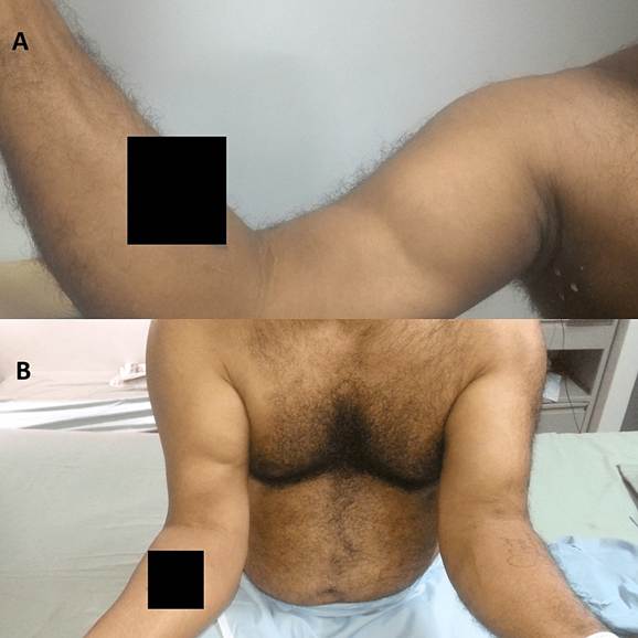
FIGURE 1: (A) Reverse Popeye sign. (B) Comparison of the distal biceps tendon rupture in the right arm with the normal left arm
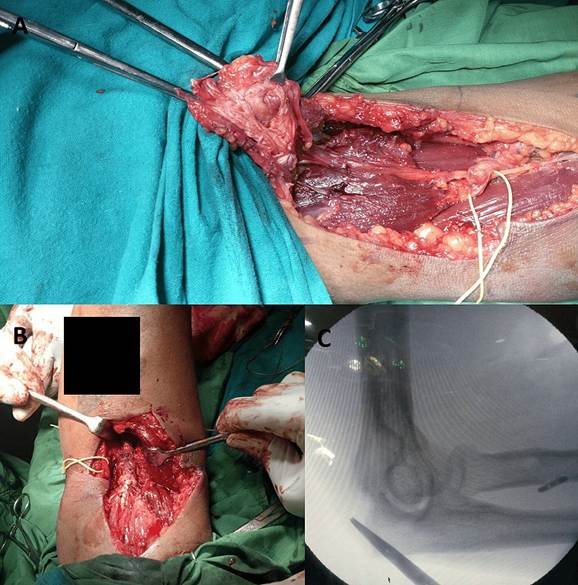
FIGURE 2: (A) Sagittal MRI image showing distal biceps tendon rupture.
(B) Axial MRI image showing distal biceps tendon rupture
The patient underwent an open anterior distal biceps tendon repair using a modified Henry’s approach. A 12-cm incision provided access to the biceps tendon and radial tuberosity. Safeguarding vital structures like the median nerve, brachial artery, and lateral antebrachial cutaneous nerve was a priority during dissection. The biceps tendon had retracted 8 cm with fraying of the distal ends (Figure 3A). A cable grafting technique was employed, utilizing the ipsilateral flexor carpi radialis, reinforced with fiber tape, to bridge the gap and ensure tension (Figure 3B, 3C).
FIGURE 3: (A) Intraoperative image showing distal biceps rupture. (B) Distal biceps repair performed with autograft augmentation. (C) Image of the arm showing tightrope attachment
Following the surgery, the patient’s elbow was immobilized at an angle of 60 degrees of flexion using a plaster of Paris slab. Subsequently, a range of motion (ROM) elbow brace was applied, featuring an extension stopper. This brace allowed controlled and gradual increases in the ROM to prevent the tendon from pulling out. During the postoperative period, activities that could potentially apply excessive force to the repaired tendon, such as forceful supination and weight loading, were avoided.
Around six weeks post-surgery, resistance exercises were initiated, and the patient regained full ROM in the affected arm. At the six-month, one-year, and five-year follow-up marks, the patient demonstrated grade 5 biceps power (Figure 4).
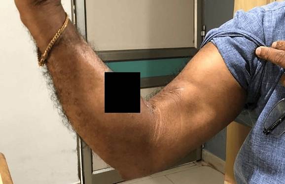
FIGURE 4: Postoperative image at six-month follow-up
Discussion
When choosing a nonoperative treatment pathway, it is essential to consider the primary role of the biceps muscle in supination and its secondary involvement in flexion. In such cases, it is important to anticipate a significant reduction in functional strength, with an approximate 50% decrement in supination capability, a 30% diminution in flexion capacity, and a 15% decline in grip strength [3].
2
The biceps tendon, with short and long heads, attaches uniquely to the radius – the short head (60 mm ) and
2
the long head (48 mm ) – posteriorly or ulnarly. Surgical repair focuses on precise reattachment [4]. The distal biceps tendon’s insertion zone is divided into three significant vascular zones. The proximal segment relies on brachial artery branches; transitionally, the recurrent branch of the posterior interosseous artery plays a crucial role, with an avascular transitional region susceptible to injury. Full forearm pronation leads to impingement at the proximal radioulnar joint, elevating injury risk by nearly 50% [5,6]. The complex anatomy and biomechanical factors form the basis of distal biceps tendon injuries [4]. Precise biceps tendon reattachment is crucial for effective treatment, especially in challenging cases with persistent injuries or tough scar tissue. Grafts, like hamstring, flexor carpi radialis, palmaris longus, and fascia lata autografts, are well-explored options. Additionally, allografts such as the Achilles tendon are studied as credible alternatives for challenging cases [7].
The surgical approach has to be preplanned, with dual-incision techniques offering augmented exposure alongside heightened concerns regarding heterotopic ossification pathological processes characterized by anomalous bone formation within soft tissues. Conversely, the single-incision approach garners favor in many clinical scenarios owing to its potential to attenuate the risk of ossification, especially when juxtaposed against methodologies such as Morrey’s modified Boyd and Anderson’s double-incision technique. While the single-incision technique does carry an elevated likelihood of posterior interosseous nerve injury, employing hypersupination of the forearm emerges as a strategic mitigatory measure. Strategic adoption of endobuttons emerges as a discerning choice, not only due to its documented superior biomechanical robustness but also due to its demonstrated propensity for minimizing mechanical failure.
Consideration of these factors augments the overall durability and resilience of the repair process [8].
The endoscopic repair method is gaining attention, offering cosmetic benefits despite challenges like brachial vessel injury and nerve issues, especially in altered cubital fossa anatomy. While effective for graftfree reconstructions with quick healing, it may be less suitable for tough scar tissue and not ideal for cases with dense scar tissue. Balancing surgery and cosmetic preferences provides a precise view but may not be as effective when grafts are required [9].
Postoperative rehabilitation spans six months, beginning with splinting and gradually progressing to extension restoration. Two weeks of immobilization was done. During the three- to six-week period, activities ranged from flexion arcs of 60 to 120 degrees with assisted movements. At six weeks, incorporating resistance exercises becomes pivotal, catalyzing methodical muscular strength restoration, culminating in unrestricted activity endorsement at six months [10].
Conclusions
Distal biceps injuries, a rarity in orthopedic clinical practice, require a tailored decision-making approach to optimize functional outcomes for patients. The success of outcomes is contingent upon considerations such as heterotopic ossification and neurovascular damage, necessitating careful evaluation of benefits on a case-by-case basis, especially in chronic injury scenarios. In instances of chronic retracted tears, the discussion further encompasses the imperative consideration of graft utilization to enhance tissue repair and restore optimal function.
Additional Information
Author Contributions
All authors have reviewed the final version to be published and agreed to be accountable for all aspects of the work.
Concept and design: Prince Shanavas Khan, Abhiram Kannan, Ayyappan V. Nair, Azaid Sait
Acquisition, analysis, or interpretation of data: Prince Shanavas Khan, Aebel Raju, Abhiram Kannan,
Ayyappan V. Nair, Yonsik Yoo, Jimmy J. Meleppuram
Drafting of the manuscript: Prince Shanavas Khan, Aebel Raju, Abhiram Kannan, Yonsik Yoo, Azaid Sait
Critical review of the manuscript for important intellectual content: Prince Shanavas Khan, Aebel
Raju, Abhiram Kannan, Ayyappan V. Nair, Azaid Sait, Jimmy J. Meleppuram
Supervision: Prince Shanavas Khan, Aebel Raju, Abhiram Kannan, Ayyappan V. Nair, Yonsik Yoo, Azaid
Sait, Jimmy J. Meleppuram
Disclosures
Human subjects: Consent was obtained or waived by all participants in this study. Conflicts of interest: In compliance with the ICMJE uniform disclosure form, all authors declare the following: Payment/services info: All authors have declared that no financial support was received from any organization for the submitted work. Financial relationships: All authors have declared that they have no financial relationships at present or within the previous three years with any organizations that might have an interest in the submitted work. Other relationships: All authors have declared that there are no other relationships or activities that could appear to have influenced the submitted work.
References
- Hsu D, Anand P, Mabrouk A, Chang KV: Biceps tendon rupture. StatPearls [Internet]. StatPearls Publishing,
Treasure Island (FL); 2024.
- Schmidt CC, Styron JF, Lin EA, Brown BT: Distal biceps tendon anatomic repair . JBJS Essent Surg Tech.
2017, 7:e32. 10.2106/JBJS.ST.16.00057
- Athwal GS, Steinmann SP, Rispoli DM: The distal biceps tendon: footprint and relevant clinical anatomy . J
Hand Surg Am. 2007, 32:1225-9. 10.1016/j.jhsa.2007.05.027
Elbow Surg. 1995, 4:149-56. 10.1016/s1058-2746(05)80044-8
- Bicos J: Biomechanics and anatomy of the proximal biceps tendon . Sports Med Arthrosc Rev. 2008, 16:111-
- 10.1097/JSA.0b013e31818247a1
- Ribeiro LM, Almeida Neto JI, Belangero PS, Pochini AC, Andreoli CV, Ejnisman B: Reconstruction of the distal biceps tendon using semitendinosus grafting: description of the technique. Rev Bras Ortop. 2018,
53:651-5. 10.1016/j.rboe.2018.07.008
- El-Hawary R, Macdermid JC, Faber KJ, Patterson SD, King GJ: Distal biceps tendon repair: comparison of surgical techniques. J Hand Surg Am. 2003, 28:496-502. 10.1053/jhsu.2003.50081
- Bhatia DN: Endoscopic repair of acute and chronic retracted distal biceps ruptures . J Hand Surg Am. 2016,
41:e501-7. 10.1016/j.jhsa.2016.09.008
- Srinivasan RC, Pederson WC, Morrey BF: Distal biceps tendon repair and reconstruction . J Hand Surg Am.
2020, 45:48-56. 10.1016/j.jhsa.2019.09.014