

Alan M. Hirahara, M.D., F.R.C.S.C., and Wyatt J. Andersen, A.T.C.
- Journal of ISAKOS – 2023
LINK: https://www.jisakos.com/article/S2059-7754(24)00042-7/fulltext
Partial articular supraspinatus tendon avulsion (PASTA) lesions present a surgical challenge, particularly in young, active patients, as they disrupt tendon integrity and risk tear progression. Transtendinous repair preserves intact bursal fibers, maintains tendon length, and enhances healing potential compared to tear completion and repair, especially for Ellman Grade 3 lesions and failed conservative cases.
Our described technique employs progressive dilation and anchor drill sleeve insertion for medial row anchor placement, with percutaneous spinal needles used for precise suture shuttling. Locking sliding knots compress the medial row repair, followed by lateral row fixation in a crisscross configuration, optimizing footprint coverage and stability. This method minimizes tendon disruption, facilitates bone-to-tendon healing, and accelerates rehabilitation.
Indicated for tears involving >50% tendon thickness or >6 mm footprint exposure, the transtendinous technique is technically demanding but preserves tendon quality and reduces retear risk. Outcomes in our series demonstrated significant functional improvement and successful return to sports at 12 months. Limitations include restricted visualization and technical difficulty, requiring surgeon expertise. Future refinements should address anchor placement optimization and tension assessment to further improve outcomes. Transtendinous repair remains a valuable option for young patients with PASTA lesions, offering durable restoration of shoulder function.
| A |
rthroscopic treatment of PASTA (partial articular supraspinatus tendon avulsion) lesions remains at the center of a great deal of debate among surgeons.1-9 Tears of greater than 50% thickness are best remedied by repair, whereas tears of less than 50% thickness are treated with subacromial decompression and debridement alone.1,3,5,6,9 Two commonly used methods are the transtendon technique and the “takedown” procedure, in which a partial-thickness tear is completed with an ensuing full-thickness repair. Both have been shown to have comparable
clinical outcomes,1,3,5,6 but they are biomechanically different.9-14 These procedures are technically difficult and have shortcomings, creating the need for a simple, easily reproducible repair technique.
Partial-thickness rotator cuff tears can be classified as intratendinous, bursal sided, or articular sided. In their
study analyzing rotator cuff footprint lesions, Schaeffeler et al.15 found that approximately 50% were articularsided tendon tears and that most of these lesions involved the anterior supraspinatus tendon. Studies have shown that restoration of the tendon footprint at the greatertuberosityisanimportantfactorintheoutcomeof rotator cuff repairs.10-14 Adequately restoring this repair site is believed to improve the biological and healing components of the repair and thereby produce more favorable outcomes.9-14 The PASTA bridge sufficiently re-creates the footprint. We present our technique for the PASTA bridgedan arthroscopic, percutaneous method of repair for PASTA lesions.
Surgical Technique
Patient Setup and Preparation
The patient is brought to the operating room and placed in the beach-chair position under general anesthesia. A diagnostic arthroscopy is performed initially to determine tissue integrity and the presence of other pathologies. Before the repair, the exposed footprint at the greater tuberosity from the PASTA lesion should be debrided down to bleeding bone in preparation for the procedure (Fig 1).
Medial-Row Preparation and Anchor Placement
| Arthroscopy Techniques, Vol 6, No 5 (October), 2017: pp e1645-e1652 e1645 |
A 17-gauge spinal needle is used as a guide to determine anchor placement at the anterior aspect of the medial row while being viewed arthroscopically
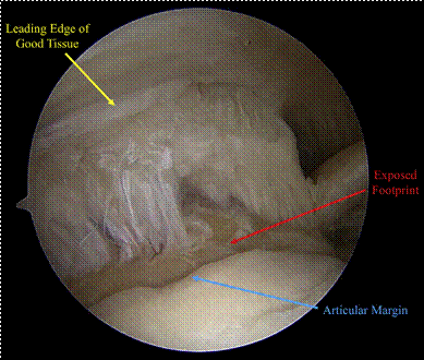
Fig 1. With the patient placed in the beach-chair position, the PASTA (partial articular supraspinatus tendon avulsion) lesion and the articular margin (blue arrow) are seen through a standard posterior portal in the left shoulder. The leading edge of good supraspinatus tendon tissue (yellow arrow) should be identified and used in the repair. The repair should reattach the supraspinatus tendon to the exposed footprint (red arrow) at the greater tuberosity.
through a posterior portal (Video 1, Fig 2). Use of the spinal needle ensures the surgeon is using suitable tissue at its leading edge and the needle is at the appropriate entry area and angle. This safeguards against damaging the surrounding structures or the tendon itself because the spinal needle will cause minimal harm if passed blindly multiple times. When
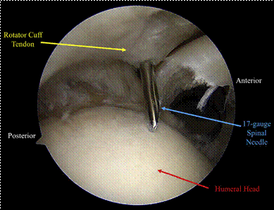
Fig 2. With the patient placed in the beach-chair position and viewing from the posterior portal in the left shoulder, the humeral head (red arrow) and rotator cuff tissue (yellow arrow) can be identified. A 17-gauge spinal needle (blue arrow) can be seen passing through the leading edge of the good rotator cuff tissue for localization of the appropriate entry and placement of sutures through the rotator cuff.
Table 1. Pearls and Pitfalls
Pearls
Use cannulas to ensure there are no soft-tissue bridges when passing sutures.
Note that progressive dilation of the entry hole limits residual damage.
Place anchors at the articular margin for supraspinatus repair.
Make sure to pass the solid white portion of the TigerWire tail back onto itself for knotless anchors.
Pitfalls
Stay away from the musculotendinous junction and tear site when placing the spinal needle.
Bear in mind that posterior anchor placements should follow
anatomic attachment sites, not the articular margin, to avoid constraining the shoulder.
Note that the anchors must be tested after placement.
Be aware that the horizontal mattress configuration must be tensioned so that the tendon is abutted to the articular margin.
using the spinal needle to determine suture passage placement, the surgeon is advised to stay at least 1 cm away from the musculotendinous junction and away from the partially torn tendon (Table 1). For the supraspinatus, the anchors should be placed at the articular margin, but for the infraspinatus, the anchors should follow the normal attachment, leaving a bare spot. The first pass is usually for the anterior-most anchor. Once established, the inner trocar of the spinal needle is replaced with a nitinol wire (Fig 3). A No. 11 blade is used to make a small percutaneous incision. A 2.4-mm Portal Dilator (Arthrex, Naples, FL) is used to dilate the portal over the nitinol wire (Fig 4). The nitinol wire is removed. In this technique a half-pipe speardas opposed to a full speardreplaces the dilator and is used to guide the punch for creation of the
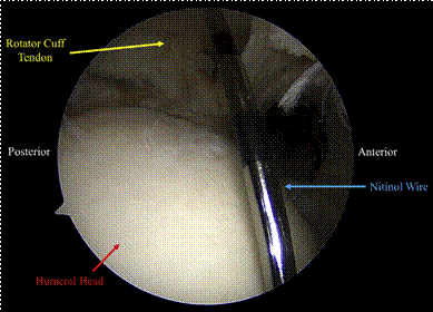
Fig 3. With the patient placed in the beach-chair position and viewing from the posterior portal in the left shoulder, the nitinol wire (blue arrow) can be seen going through the rotator cuff tissue (yellow arrow) to the humeral head (red arrow). The initial hole is made with a spinal needle, and the nitinol wire replaces the spinal needle.
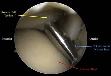
Fig 4. With the patient placed in the beach-chair position and viewing from the posterior portal in the left shoulder, the 2.4mm portal dilation tube (blue arrow) can be seen going through the rotator cuff tendon (yellow arrow) for progressive dilation for eventual anchor placement. The portal dilation tube acts as a guide for the half-pipe spear used to place the Knotless Corkscrew anchor.
anchor sockets (Fig 5). The half-pipe spear is used so that a smaller hole is made through the rotator cuff. The spear can be moved as necessary to determine optimal anterior anchor placement, usually next to the bicipital groove at the articular margin. Because the greater tuberosity is made of soft, cancellous bone, a punch is used to create a socket (Fig 6). A 3.9-mm Knotless Corkscrew anchor (Arthrex) can be used in this technique (Fig 7). The anchor is placed into the socket.
For placement of the anchor at the posterior aspect of the medial row, the procedure is the same as that of the anchor at the anterior aspect. A 17-gauge spinal needle is used to identify the correct location and leading edge of the appropriate tissue. A nitinol wire replaces the spinal needle. A No. 11 blade is used to make a small percutaneous incision at the nitinol wire. A dilator is
Fig 5. With the patient placed in the beach-chair position and viewing from the posterior portal in the left shoulder, a halfpipe spear (blue arrow) is passed through the rotator cuff tendon (yellow arrow). A half-pipe spear is used to limit the size of the hole going through the rotator cuff.
exchanged for the wire and then removed when the half-pipe spear is introduced. The spear is adjusted as necessary to ensure optimal anchor placement; then, a punch is used to create a posterior socket. A 3.9-mm Knotless Corkscrew anchor is placed into the socket.
Medial-Row Mattress Fixation
With the medial-row anchors placed, the arthroscope is moved to the subacromial space. The attached TigerWire (Arthrex) from the posterior anchor (Fig 8) and the FiberLink (Arthrex), the looped passing suture, from the anterior anchor (Fig 9) are collected with a grasper and pulled through an 8 3emm PassPort cannula (Arthrex). The TigerWire used in this technique has a white section and a black-and-white striped section (Fig 10). These areas denote changes in diameter, with the solid white area being a smaller diameter. The TigerWire is passed through the FiberLink almost
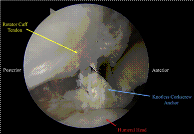
Fig 7. With the patient placed in the beach-chair position and viewing from the posterior portal in the left shoulder, a Knotless Corkscrew anchor (blue arrow) is placed into the anterior socket. The anchor is passed through the half-pipe spear into the socket that was created by the punch.
back to the black-and-white striped section so that there is plenty of suture to shuttle through the knotless anchor (Fig 11). The suture end of the FiberLink is then pulled from the anterior anchor to pass the TigerWire until it is snug but not tensioned.
The same process is repeated for the posterior anchor. The FiberLink from the posterior anchor and the TigerWire from the anterior anchor are gathered and pulled with a grasper through the 8 3emm PassPort cannula. The TigerWire is passed through the FiberLink. The suture end at the posterior anchor is pulled to pass the TigerWire through the knotless anchor. The 2 suture ends can then be pulled alternately to tension
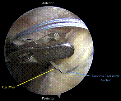
Fig 8. With the patient placed in the beach-chair position and viewing the subacromial space from the lateral portal in the left shoulder, a grasper is introduced to pass the TigerWire through the anterolateral portal. In this image, the TigerWire is from the posterior Knotless Corkscrew anchor.
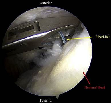
Fig 9. With the patient placed in the beach-chair position and viewing the subacromial space from the lateral portal in the left shoulder, a grasper is introduced to pass the FiberLink from the anterior Corkscrew anchor through the anterolateral portal. The FiberLink is a looped passing suture that is used to pass the TigerWire from the opposing anchor to create a horizontal mattress.
the horizontal mattress (Fig 12). Visualization with the arthroscope can help to ensure the rotator cuff tissue is abutted and tensioned to the articular margin appropriately. The construct can be tested and re-tensioned as necessary. This completes the medial-row anchor construct.
Lateral Anchor Fixation
The remaining sutures are gathered and pulled through the lateral portal (Fig 13). A Vented SwiveLock (Arthrex) is then attached to the suture. A standard punch is used to create a lateral socket. The SwiveLock is introduced and fixed into position at the lateral aspect of the construct (Fig 14). The remaining suture can be cut (Fig 15), completing the repair (Fig 16).
Rehabilitation
The initial phase of rehabilitation after the PASTA bridge begins within 1 week of surgery to commence early passive range of motion. A sling is a requirement for the first 3 to 4 weeks for protection of the repair and for comfort. By 6 weeks, full active range of motion should be achieved, and light rotator cuff and scapular strengthening can begin. By 12 weeks, the patient can begin moderate lifting, weight training, and plyometrics. Return to play and participation in a throwing program begin at 5 months. Return to nonthrowing sports occurs at approximately 8 months. Overhead athletes can return to play between 9 and 12 months.
Discussion
Partial-thickness rotator cuff tears are a prevalent injury, yet there is no consensus on the optimal repair technique. Although many procedures have been developed, 2 traditional methods are transtendon repair and the takedown method of propagating a partial tear to completion with a subsequent full-thickness repair. The transtendon repair is technically very difficult and could result in damage to the surrounding tissues while trying to introduce the anchor into the joint blindly or trying to create a blind hole in the tendon with a scalpel or trocar. Furthermore, the takedown method takes a partial tear into a full tear. If the repair fails, the patient is left with a full-thickness tear that could be more debilitating and painful. Repaired partial cuff tears also exhibit significantly improved biomechanical properties when compared with full-thickness repairs.12-14 Some surgeons have been reluctant to perform these surgical procedures because of the risks and technical difficulty inherent to the procedures. Although reports on the 2 aforementioned procedures have produced adequate outcomes,1,3,5,6 the percutaneous nature of the PASTA bridge makes it a superior technique
(Table 2).
Re-creating the rotator cuff footprint at the greater tuberosity is the primary goal of arthroscopic rotator cuff repair. Providing greater contact at this important site has the potential to improve healing.9-11,13,14 Double-row repair techniques have been shown to provide greater pressure patterns, increase contact pressures, and restore larger footprints than single-row procedures.12,13 The PASTA bridge re-establishes this tendon-bone interface in a similar manner. Placing anchors laterally in conjunction with a medial row has led to superior outcomes resulting from an increase in the repair site contact area.10 The PASTA bridge uses a lateral anchor to take all the stress off the construct being pulled medially and only using the medial anchors as pivot points. Double-row techniques have been argued to be superior to single-row procedures by
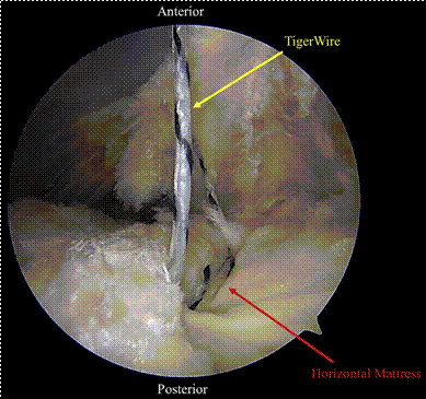
Fig 12. Withthepatientplacedinthebeach-chairposition and viewing the subacromial space through the lateral portal in the left shoulder, the medial horizontal mattress can be visualized. After the TigerWire is shuttled through the opposing anchor through the FiberLink at each anchor, the medial horizontal mattress is created. This completes the medial portion of the PASTA(partialarticularsupraspinatustendonavulsion)bridge.
increasing the number of fixation points.9 A larger collection of fixation points reduces stress and load at each suture point, making the overall repair more robust and reliable.9
Anchor placement during the PASTA bridge is performed with a spinal needle. The spinal needle allows the surgeon to choose his or her site of entry and can be placed or moved intraoperatively without damaging surrounding tissue. This is in contrast to other techniques that use a scalpel or spear as their method of entry, which could cause iatrogenic damage to surrounding tissues. For this reason, many surgeons have historically been hesitant to repair partial cuff tears of less than 50% thickness. This technique makes smaller tears more amenable to repair.
For repairs that require multiple anchors, placement is difficult and cumbersome in traditional methods. The PASTA bridge’s percutaneous technique is safe and reliable for single- or multiple-anchor repairs. The angle of anchor placement varies by patient and can be optimized by using the spinal needle. Because of its unobtrusive nature, the PASTA bridge can be used for tears of virtually any size or thickness. Ideally, the leading edge of the good tissue should be used. If the anchors are placed too medially near the musculotendinous margin, tear through may occur. If the anchors are placed into the PASTA defect, the tissue may not hold. If a delamination tear is present, the PASTA bridge can reliably incorporate this tissue into
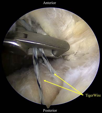
Fig 13. Withthe patientplaced inthebeach-chair position and viewing the subacromial space through the lateral portal in the left shoulder, a grasper can be viewed pulling the remaining TigerWire sutures through an anterolateral portal. After the medial-row construct is completed, the TigerWire sutures are pulled through a lateral portal to be attached to a Vented SwiveLock for lateral fixation and completion of the PASTA (partial articular supraspinatus tendon avulsion) bridge.
the repair. Although this procedure is aimed toward PASTA lesions, the technique can be used in a variety of settings, including supraspinatus tears, infraspinatus
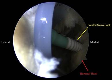
Fig 14. With the patient placed in the beach-chair position and viewing the subacromial space through the lateral portal in the left shoulder, a Vented SwiveLock is placed into the lateral socket. The Vented SwiveLock is attached exteriorly to the TigerWire sutures from the medial-row anchors for lateral fixation. The lateral anchor assists in distributing force across the repair, reducing load at each anchor point.
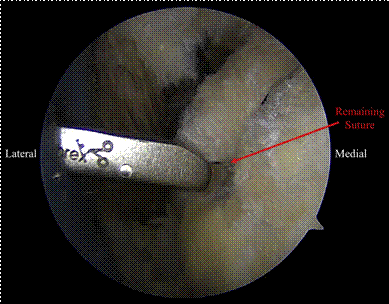
Fig 15. Withthepatientplacedinthebeach-chairposition and viewing the subacromial space through the lateral portal in the left shoulder, the Vented SwiveLock is fully seated to the bone and the remaining sutures are cut flush with the anchor. This avoids knot stacks that have the potential to cause iatrogenic defects.
tears, subscapularis tears, and remplissage. The PASTA bridge is a reliable procedure that is appropriate for surgeons of all experience levels. Given its technical ease and percutaneous nature, the PASTA bridge should be considered when treating PASTA lesions.
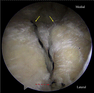
Fig 16. With the patient placed in the beach-chair position and viewing the subacromial space through the lateral portal in the left shoulder, the final construct can be visualized. The TigerWire sutures (solid yellow arrows) from the anterior and posterior knotless anchors of the medial row are shown coming through the rotator cuff to be fixed laterally (dashed red arrow) to a Vented SwiveLock anchor in the bone.
Table 2. Advantages and Disadvantages
Advantages
Spinal needle used for accurate anchor placement
Half-pipe spear minimizes hole through tendon
Repairs tears of any size
Easy to place multiple anchors
Threaded anchor for better fixation
Knotless anchors and fixation
Percutaneous procedure
Easily reproducible
Disadvantages
Good suture management needed so as not to cross sutures when creating knotless construct
Surgeon must be sure to use appropriate sutures from correct anchors when establishing medial row
References
- Burkhart SS, Danaceau SM, Pearce CE Jr. Arthroscopic rotator cuff repair: Analysis of results by tear size and by repair techniquedMargin convergence versus direct tendon-to-bone repair. Arthroscopy 2001;17:905-912.
- Budoff JE, Rodin D, Ochiai D, Nirschl RP. Arthroscopic rotator cuff debridement without decompression for the treatment of tendinosis. Arthroscopy 2005;21:1081-1089.
- Castagna A, Delle Rose G, Conti M, Snyder SJ, Borroni M, Garfalo R. Predictive factors of subtle residual shoulder symptoms after transtendinous arthroscopic cuff repair: A clinical study. Am J Sports Med 2009;37:103-108.
- Cordasco FA, Backer M, Craig EV, Klein D,
Warren RF. The partial-thickness rotator cuff tear: Is acromioplasty without repair sufficient? Am J Sports Med 2002;30:257-260.
- Ide J, Maeda S, Takagi K. Arthroscopic transtendon repair of partial-thickness articular-side tears of the rotator cuff: Anatomical and clinical study. Am J Sports Med 2005;33: 1672-1679.
- Kamath G, Galatz LM, Keener JD, Teefey S, Middleton W, Yamaguchi K. Tendon integrity and functional outcome after arthroscopic repair of highgrade partial-thickness supraspinatus tears. J Bone Joint Surg Am 2009;91:1055-1062.
- Kartus J, Kartus C, Rostgård-Christensen L, Sernet N, Read J, Perko M. Long-term clinical and ultrasound evaluation after arthroscopic acromioplasty in patients with partial rotator cuff tears. Arthroscopy 2006;22:44-49.
- Liem D, Alci S, Dedy N, Steinbeck J, Marquardt B, Möllenhoff G. Clinical and structural results of partial supraspinatus tears treated by subacromial decompression without repair. Knee Surg Sports Traumatol Arthrosc 2008;16:967-972.
- Lo IKY, Burkhart SS. Double-row arthroscopic rotator cuff repair: Re-establishing the footprint of the rotator cuff. Arthroscopy 2003;19:1035-1042.
- Apreleva M, Özbaydar M, Fitzgibbons PG, Warner JJP. Rotator cuff tears: The effect of the reconstruction method on three-dimensional repair site area. Arthroscopy 2002;18:519-526.
- Baums MH, Spahn G, Steckel H, Fischer A, Schultz W, Klinger HM. Comparative evaluation of the tendon-bone interface contact pressure in different single- versus double-row suture anchor repair techniques. Knee Surg Sports Traumatol Arthrosc 2009;17:1466-1472.
- Gonzalez-Lomas G, Kippe MA, Brown GD, et al. In situ transtendon repair outperforms tear completion and repair for partial articular-sided supraspinatus tendon tears. J Shoulder Elbow Surg 2008;17:722-728.
- Mazzocca AG, Millett PJ, Guanche CA, Santangelo SA, ArcieroRA.Arthroscopicsingle-rowversusdouble-rowsuture anchorrotatorcuffrepair.AmJSportsMed2005;33:1861-1868.
- Mazzocca AD, Rincon LM, O’Connor RW, et al. Intraarticular partial-thickness rotator cuff tears: Analysis of injured and repaired strain behavior. Am J Sports Med 2008;36:110-116.
- Schaeffeler C, Mueller D, Kirchhoff C, Wolf P, Rummeny EJ, Woertler K. Tears at the rotator cuff footprint: Prevalence and imaging characteristics in 305 MR arthrograms of the shoulder. Eur Radiol 2011;21: 1477-1484.