

Arun Mundakkal, D.N.B. Ortho., Mohamed Mansoof Kanakkayil, D.N.B. Ortho.,
Radhesh Nambiar, D.Ortho., Azad Sait, D.Ortho., M.S.Ortho.,
Ayyappan V. Nair, D.Ortho., D.N.B.Ortho., Yon-Sik Yoo, M.D., Ph.D., and Prince Shanavas Khan, D.Ortho., M.S. Ortho.
- Arthroscopy Techniques – 2022
LINK: https://www.arthroscopytechniques.org/article/S2212-6287(22)00258-4/fulltext
The subscapularis muscle, the strongest rotator cuff muscle, plays a vital role in maintaining balanced glenohumeral joint forces. Tears of the subscapularis tendon compromise shoulder stability and function but are difficult to diagnose and repair arthroscopically due to limited visualization. This Technical Note describes the use of an anterolateral viewing portal combined with the lasso‑loop stitch technique to improve visualization, tendon mobilization, and repair strength.
Under general and interscalene-block anesthesia with the patient in lateral decubitus position, diagnostic arthroscopy is performed via a posterior portal. A capsulotomy at the supraspinatus edge allows creation of the anterolateral viewing portal, providing a direct view of the subscapularis tendon. Extensive release of the tendon is performed under visualization while safeguarding the axillary nerve. The tendon footprint is prepared, and a double‑loaded suture anchor is placed in the lesser tuberosity. The inferior tendon portion is repaired with a mattress stitch, while the superior portion is secured with a lasso‑loop stitch for enhanced tissue grip and repair strength.
This method provides superior visualization, facilitates extensive tendon release, and achieves secure repair of retracted subscapularis tears, improving functional outcomes. The technique is technically demanding but reproducible and advantageous for difficult subscapularis repairs.
| S |
ubscapularis tendon tears were first described by recognition of partial tears is under-reported.4 The Smith and recognized by Codman.1 Hauser2 re- incidence of subscapularis tears was shown to be be-
5
| ported the first case of subscapularis tendon repair in 1954. Various open techniques for the repair of the subscapularis tendon tears were described. Burkhart and Tehrany3 published the first arthroscopic technique of repair of subscapularis tendon tear in 2002. With improvements in clinical examination and radiographic imaging studies, our ability to accurately diagnose subscapularis tears has improved, but the | tween 3% and 13% by autopsy and cadaveric studies. Subscapularis tears are hard to access, and they require advanced surgical techniques.6 Various repair techniques of torn subscapularis tendon have been described recently. Most of the recent literature has studied the outcome of arthroscopic subscapularis tendon repair using various techniques. Most of the studies used single-row/double-row repair or mattress sutures for arthroscopic subscapularis |
tendon repair.
In 2006, a new suture technique was introduced by Lafosse et al.6 to improve the tissue gripdthe lasso loop stitch, which initially was described for the reattachment of the rotator cuff. It was also described for Bankart repair and biceps tenodesis.7 In 2010, Lafosse et al.6 described the arthroscopic repair of subscapularis tendon tear using the lasso-loop technique and mattress sutures and demonstrated good clinical outcome.
The anterolateral portal offers better visualization of the articular and bursal surface of the subscapularis tendon and aids in release of the retracted subscapularis tendon tears. In this Technical Note, we present our preferred technique of subscapularis repairdarthroscopic repair of subscapularis tendon using the lassoloop technique through anterolateral viewing portal.
Arthroscopy Techniques, Vol 12, No 1 (January), 2023: pp e83-e89 e83
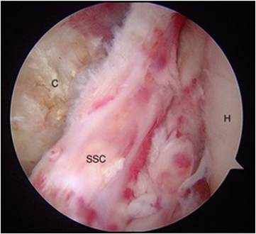
Fig 1. Patient shown in the lateral decubitus position. Posterior viewing portal established. Diagnostic arthroscopy through the posterior viewing portal showing Subscapularis tear. (C, coracoid; H, head of humerus; SSC, subscapularis.)
Surgical Technique (With Video Illustration)
Patient Positioning
Surgery is performed under general anesthesia and interscalene-block anesthesia. The patient can be positioned in either the beach-chair or lateral decubitus position. We position the patient in lateral decubitus position with the arm in 30 abduction and 20 flexion and traction. After sterile preparation and draping, bony landmarks are outlined.
Portal Placement
We use 3 portals: the standard posterior viewing portal, the anterior working portal, and an anterolateral viewing portal. A standard posterior portal is used to perform a routine diagnostic arthroscopy of the glenohumeral joint (Fig 1). The subscapularis tear is arthroscopically classified according to the Lafosse et al.6 classification to decide the treatment. The anterolateral portal is established at the leading edge of the supraspinatus tendon. A spinal needle is inserted into the glenohumeral joint at the leading edge of the supraspinatus tendon. A no. 11 blade is advanced collinear to the spinal needle into the glenohumeral joint. Make a capsulotomy with the no. 11 blade at the leading edge of the supraspinatus. Using a switching stick, a trocar cannula is inserted and anterolateral viewing portal is established (Fig. 2).
Subscapularis Release
Anterolateral portal offers better visualization of the articular and bursal surface of the subscapularis tendon. The retracted subscapularis tendon can be released extensively on the articular and bursal surface by visualizing through the anterolateral viewing portal (Fig 3). At most, care shoulder be taken to avoid injury to the axillary nerve at the bursal surface of the subscapularis tendon. The radiofrequency ablator probe should always be pointing upwards so that the tip can be visualized. A traction stitch placed in the tendon can assist in mobilization of the tendon, if the tendon is severely retracted. The tendon can be extensively released to achieve full excursion to its footprint on the lesser tuberosity to allow for a tension free repair.
Subscapularis Repair: The Lasso-Loop Technique
The lesser tuberosity is debrided and decorticated with radiofrequency ablator and burr before the placement of anchor (Fig 4). A double-loaded anchor (CEPTRE 4.8-mm PEEK suture anchor; Sironix Renewables, Seattle, WA) is placed into the lesser tuberosity at the subscapularis tendon footprint (Fig 5). One suture of the double-loaded anchor is employed for mattress suture in the inferior aspect of the tendon (Fig 6). The 2 limbs of the suture are retrieved using a suture shuttling device (Rhino suture passer, Arthrex, Naples, FL) through the inferior part of the tendon before creating the lasso-loop stitch in the superior aspect using the other suture (Fig 7). To create a lasso-loop stitch, select one limb of the suture (Fig 8) and retrieve a loop of the suture through the tendon using the suture shuttling device/bird beak to create a loop (Fig 9). Retrieve the
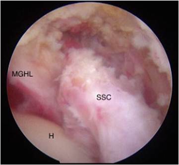
Fig 2. Patient shown in the lateral decubitus position. Viewing through the posterior portal, anterolateral portal is established at the leading edge of the supraspinatus tendon. Using a switching stick, the trocar cannula is inserted in the anterolateral portal and arthroscope inserted. The anterolateral viewing portal allows better visualization of the torn subscapularis tendon. (H, head of humerus; MGHL, middle glenohumeral ligament; SSC, subscapularis.)
subscapularis.)
distal part of the same suture limb through the suture loop to form the lasso-loop stitch (Fig 10). Pull the other limb of the suture to tighten the lasso-loop stitch (Fig 11). Use an arthroscopic non-sliding knot to complete the lasso-loop stitch over the superior part of the tendon (Fig 12). The arm is placed in neutral position for proper tendon tensioning. The lower part of the tendon is repaired using vertical mattress suture over the bursal surface with the two limbs of the suture
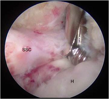
Fig 4. Patient shown in the lateral decubitus position. Viewing through the posterior portal. Working through the anterior portal. Subscapularis footprint at the lesser tuberosity is debrided and decorticated with radiofrequency ablator and burr before the placement of anchor. (H, head of humerus;
SSC, subscapularis.)
retrieved initially (Fig 13). Thus, the lasso-loop stitch secures the superior part and the vertical mattress suture secures the inferior part of the subscapularis tendon (Fig 14). Once the subscapularis repair is completed, other pathologies can be addressed.
Postoperative Rehabilitation
The operated shoulder is immobilized in a sling for 4 to 6 weeks postoperatively. Wrist and elbow motions are encouraged during the period. Passive external rotation is permitted but limited to 0 or as determined during the surgery. Forward flexion exercises are initiated once the period of immobilization ends, progressing from active-assisted to active range of motion. Deltoid and rotator cuff strengthening exercises are started at 12 weeks postoperatively, with progressive return to functional activities and sports.
Discussion
Codman1 described the concept of coordinated rotator cuff muscle activity that maintain the humeral head in glenoid fossa. Subscapularis muscle plays a significant role in active stabilization of humeral head in the glenoid fossa during external rotation and abduction.8,9 The subscapularis muscle provides the coupled balanced force across the glenohumeral joint.10 It functions by resisting the infraspinatus in axial plane and the shearing force of deltoid in the coronal
plane.10,11 When a subscapularis tear is left unrepaired, the function of the subscapularis muscle will be compromised. Furthermore, the posterosuperior rotator cuff tear repair will be difficult and less securely
repaired.12
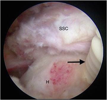
Fig 5. Patient shown in the lateral decubitus position. Viewing through the anterolateral portal and working through the anterior portal. A double loaded anchor is placed at the Subscapularis tendon footprint at the lesser tuberosity. Arrow shows the anchor inserted. (H, head of humerus; SSC,
subscapularis.)
Arthroscopy remains the gold standard for the diagnosis of subscapularis tendon tears.13 Subscapularis repair techniques are technically demanding because of difficult visualization and instrumentation. Expertise in the arthroscopic examination of the shoulder have supplemented magnetic resonance imaging in
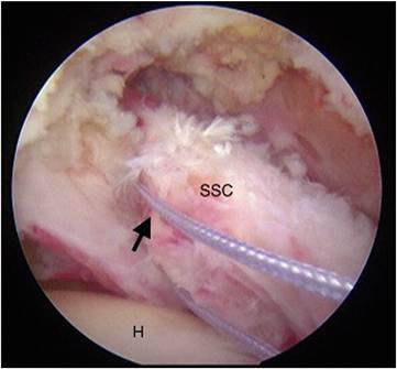
Fig 6. Patient shown in the lateral decubitus position. Viewing through the anterolateral portal and working through the anterior portal. One suture of the double-loaded anchor is selected for mattress suture in the inferior aspect of the tendon. Arrow shows the selected limb of the suture. (H, head of humerus; SSC, subscapularis.)
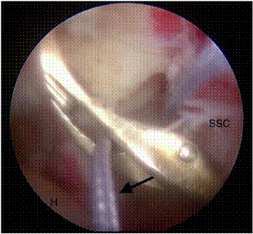
Fig 7. Patient shown in the lateral decubitus position. Viewing through the anterolateral portal and working through the anterior portal. One suture of the double-loaded anchor is selected for mattress suture. The selected limb of the suture is retrieved using a suture shuttling device or bird beak through the inferior part of the tendon. The arrow shows the selected limb of the suture. The other limb of the suture is also retrieved through the Subscapularis tendon in the same manner before creating the lasso-loop stitch. The suture is tied only after completing the lasso-loop stitch. (H, head of humerus; SSC, subscapularis.)
early and timely detection and treatment of even small tears of the subscapularis tendon. The comma sign is an arthroscopic finding indicating the presence of a
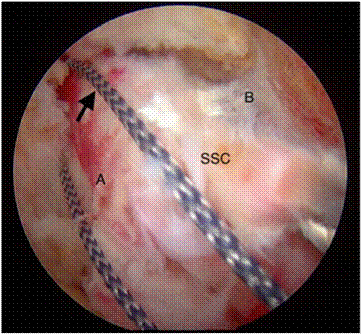
Fig 8. Patient shown in the lateral decubitus position. Viewing through the anterolateral portal and working through the anterior portal. One limb of the second suture of the double loaded anchor is selected for the lasso-loop stitch. The arrow shows the selected limb of the suture. (A, articular surface; B, bursal surface; SSC, subscapularis.)
suture. (H, head of humerus; SSC, subscapularis.)
full-thickness subscapularis tears. It represents insertional fibers of superior part of subscapularis tendon torn horizontally from residual caudal fibers, with torn insertional fibers of the superior glenohumeral ligament and coracohumeral ligament retracted superiorly and medially in a vertical orientation, forming an abnormal vertical comma-shaped band obscuring the rotator interval. The sensitivity of identifying the comma sign on magnetic resonance imaging is 63%.14 Visualization of subscapularis tendon is important to obtain an ideal anatomical repair. Most often, a 70 arthroscope may be needed for adequate visualization of the tear and the subscapularis footprint on the lesser tuberosity.15 Establishment of an anterolateral viewing portal may obviate the need for the switching of arthroscope. An anterolateral viewing portal provides excellent visualization of both the articular and bursal surface of the subscapularis. Retracted subscapularis tears can be extensively released along the articular and bursal surface through the anterolateral portal, as the neurovascular structures can be visualized better and safeguarded.
Various repair techniques of subscapularis tendon tears have been described in the literature. Of all the techniques, the lasso-loop technique described by Lafosse et al.4 provides better tissue grip and can be used especially in retracted subscapularis tears where the tendon can be reattached over the lesser tuberosity
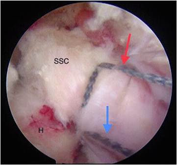
Fig 11. Patient shown in the lateral decubitus position. Viewing through the anterolateral portal and working through the anterior portal. Lasso-loop stitch is created with one limb of the suture. The second limb of the suture is pulled to tighten the lasso-loop stitch. The red arrow shows the lassoloop stitch. The blue arrow shows the second limb of the suture. (H, head of humerus; SSC, subscapularis.)
with least chances of loosening of suture. In a recent systematic review done by Xiao et al.,16 the majority of the studies used single-row repair techniques. Of all the studies described, 2 studies done by Grueninger
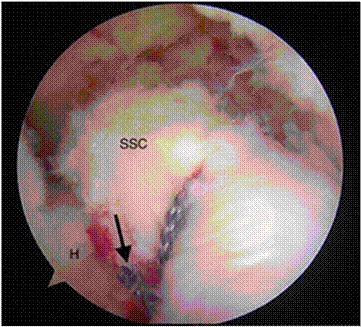
Fig 12. Patient shown in the lateral decubitus position. Viewing through the anterolateral portal and working through the anterior portal. Lasso-loop stitch is created with one limb of the suture and tightened by pulling the other limb of the suture. Tie using a non-sliding knot to complete the lasso-loop stitch over the superior part of the tendon. The arm is placed in neutral position for proper tendon tensioning. The black arrow shows the lasso-loop stitch secured with nonsliding knots. (SSC, subscapularis.)
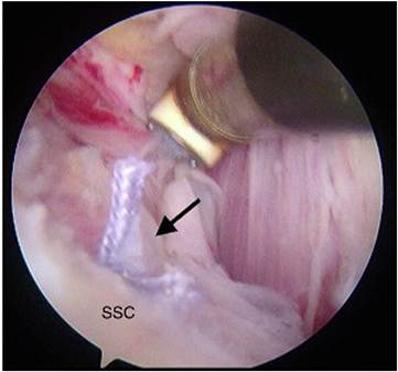
Fig 13. Patient shown in the lateral decubitus position. Viewing through the anterolateral portal and working through the anterior portal. After repairing the superior part of the subscapularis tendon using the lasso-loop technique, the lower part of the tendon is repaired using vertical mattress suture over the bursal surface with the two limbs of the suture retrieved initially. (SSC, subscapularis.) et al.17,18 used the lasso-loop stitch. Although doublerow repair techniques reported fewer retear rates, both single- and double-row repair techniques produce satisfactory outcomes clinically.16 Although technically demanding, the use of anterolateral viewing portal further simplifies the repair of subscapularis tendon tear, with better visualization of the torn tendon. The advantages and disadvantages of our techniques are
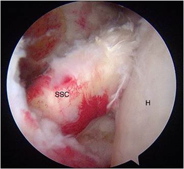
Fig 14. Patient shown in the lateral decubitus position. Viewing through the posterior portal showing repaired subscapularis tendon. (H, head of humerus; SSC, subscapularis.).
Table 1. Advantages and Disadvantages of Our Technique
| Advantages | Disadvantages |
| End on view of subscapularis tendon through the anterolateral portal Extensive release of retracted subscapularis can be done Axillary nerve injury can be avoided during release due to better visualization of the bursal surface Lasso-loop technique provides improved tissue grip and strong repair, especially in retracted tears No need of switching from a arthroscope to a 70 30 arthroscope, as the anterolateral viewing portal provides adequate visualization with a 30 arthroscope | Technically demanding Establishment of anterolateral portal requires expertise |
described in Table 1. The surgical procedure is demonstrated in Video 1.
References
- Codman EA, Peltier LF. The classic: Rupture of the supraspinatus tendon. Clin Orthop Rel Res 1990;254:3-26.
- Hauser ED. Avulsion of the tendon of the subscapularis muscle. J Bone Joint Surg 1954;36:139-141.
- Burkhart SS, Tehrany AM. Arthroscopic subscapularis tendon repair: Technique and preliminary results. Arthroscopy 2002;18:454-463.
- Lafosse L, Jost B, Reiland Y, Audebert S, Toussaint B, Gobezie RG. Structural integrity and clinical outcomes after arthroscopic repair of isolated subscapularis tears. J Bone Joint Surg Am 2007;89:1184-1193.
- Arun GR, Kumar P, Patnaik S, et al. Outcome of arthroscopic subscapularis tendon repair: Are the results improving with improved techniques and equipment? A retrospective case series. Indian J Orthop 2016;50:297-302.
- Lafosse L, Lanz U, Saintmard B, Campens C. Arthroscopic repair of subscapularis tear: Surgical technique and results. Orthop Traumatol Surg Res 2010;96:S99-108.
- Toussaint B, Schnaser E, Lafosse L, Bahurel J, Gobezie R. A new approach to improving the tissue grip of the medialrow repair in the suture-bridge technique: The “modified lasso-loop stitch. Arthroscopy 2009;25:691-695.
- Kronberg M, Németh G, Broström LA. Muscle activity and coordination in the normal shoulder. An electromyographic study. Clin Orthop 1990;257:7685.
- Perry J. Anatomy and biomechanics of the shoulder in throwing, swimming, gymnastics, and tennis. Clin Sports Med 1983;2:247-270.
- Burkhart SS. Arthroscopic treatment of massive rotator cuff tears. Clinical results and biomechanical rationale. Clin Orthop 1991;(267):45-56.
- Aluisio FV, Osbahr DC, Speer KP. Analysis of rotator cuff muscles in adult human cadaveric specimens. Am J Orthop Belle Mead NJ 2003;32:1249.
- Ticker JB, Burkhart SS. Why repair the subscapularis? A logical rationale. Arthroscopy 2011;27:1123-1128.
- Ono Y, Sakai T, Carroll MJ, Lo IK. Tears of the subscapularis tendon: A critical analysis review. JBJS Rev 2017;5(3).
- Atinga A, Dwyer T, Theodoropoulos JS, Dekirmendjian K, Naraghi AM, White LM. Preoperative magnetic resonance imaging accurately detects the arthroscopic comma sign in subscapularis tears. Arthroscopy 2021;37:3062-3069.
- Bell JP, Field LD. Combined intra-articular and extraarticular visualization for repair of a complete subscapularis tear: The “blended view” technique. Arthrosc Tech 2021;10:e1879-1882.
- Xiao M, Cohen SA, Cheung EV, Abrams GD, Freehill MT. Arthroscopic single and double row repair of isolated and combined subscapularis tears result in similar improvements in outcomes: A systematic review. Arthroscopy 2022;38:159-173.
- Grueninger P, Nikolic N, Schneider J, et al. Arthroscopic repair of traumatic isolated subscapularis tendon lesions (Lafosse type III or IV): a prospective magnetic resonance imagingecontrolled case series with 1 year of follow-up. Arthroscopy 2014;30:665-672.
- Grueninger P, Nikolic N, Schneider J, et al. Arthroscopic repair of massive cuff tears with large subscapularis tendon ruptures (Lafosse III/IV): A prospective magnetic resonance imagingecontrolled case series of 26 cases with a minimum follow-up of 1 year. Arthroscopy 2015;31: 2173-2182.