Written by: Editorial Team of Dr Ayyappan V Nair
Medically Reviewed by: Dr. Ayyappan V. Nair
Senior Consultant – Shoulder Surgery, Arthroscopy and Sports Trauma
Manipal Hospitals, Bangalore
Last Reviewed: 24th September 2026
Introduction
Rotator cuff tears (RCTs) are among the most common causes of shoulder pain and dysfunction. They affect approximately 16–34% of the general population [1], with the prevalence increasing substantially with age and reaching up to 50% in individuals older than 50 years [2].
Despite improvements in surgical techniques, fixation constructs and rehabilitation protocols, structural failure after rotator cuff repair remains an important clinical problem, particularly in large and massive tears, retracted tears, poor-quality tendon, and revision cases, says Dr Ayyappan V Nair, considered as the best shoulder surgeon in India.
Biological and mechanical augmentation has therefore gained increasing attention. Synthetic and bioinductive implants have been developed to reinforce the repair, increase tendon thickness and potentially stimulate tissue regeneration. At the same time, autologous tissues—particularly the long head of the biceps tendon (LHBT) and fascia lata—offer an attractive alternative because they provide living tissue without the requirement for a separate donor site or expensive manufactured implants.
The central question is not simply whether augmentation works, but rather:
Should we favour a manufactured scaffold or the patient’s own tissue when augmentation is required?
Synthetic Augmentation: The Current Landscape
Several commercially available implants have been developed for rotator cuff augmentation.
These include:
- REGENETEN® bioinductive implant
- BioBrace® collagen/PLLA scaffold
- Tapestry® biointegrative implant
- Integrity® hyaluronic acid/PET implant
These implants differ considerably in composition, architecture, degradation characteristics and proposed biological mechanisms.
REGENETEN®
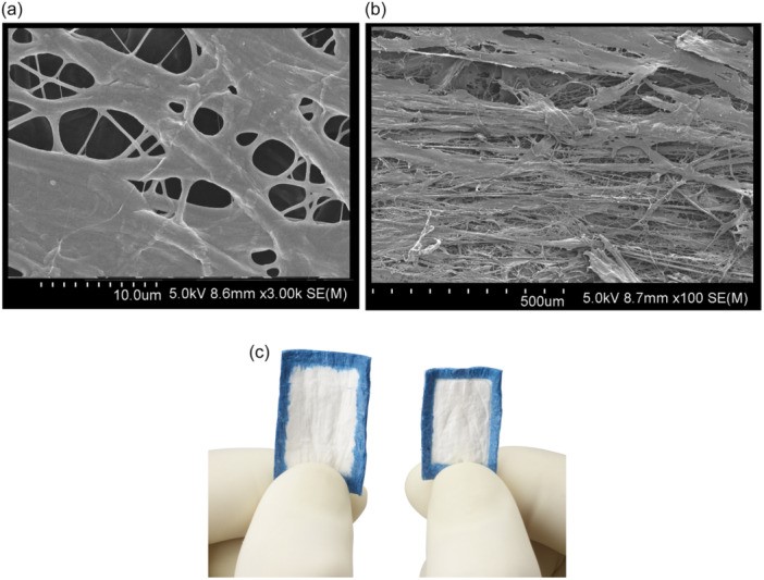
REGENETEN® is a type-I bovine collagen membrane. It was initially implanted clinically in Australia in 2012, followed by introduction into the US market in 2014 and European Union approval in 2020.
Preclinical studies demonstrated that the implant becomes populated by local tenoblasts and is progressively remodelled into tendon-like tissue. Human histological studies have similarly suggested connective-tissue formation during the first 12–24 weeks, with substantial scaffold resorption by approximately 26 weeks.
BioBrace®
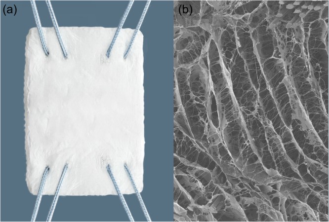
BioBrace® combines highly porous type-I bovine collagen with bioresorbable PLLA microfilaments. Its three-dimensional architecture is designed to permit tissue ingrowth while providing load-sharing mechanical support.
However, the clinical evidence remains considerably less mature than that available for REGENETEN®. The presentation cites a retrospective series of 49 large-to-massive tears reporting a 94% healing rate at two years, with reoperation and complication rates of 8% and 14%, respectively.
Tapestry®
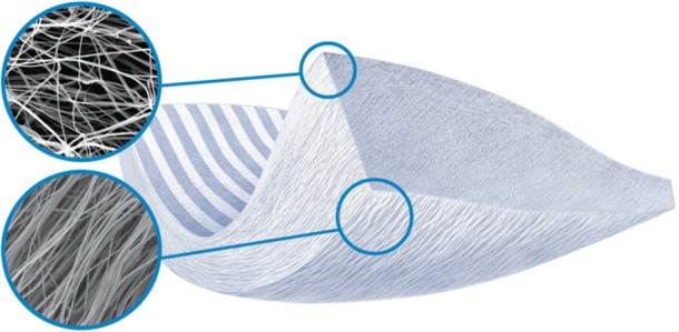
Tapestry® is designed with an aligned, porous microarchitecture intended to resemble native tendon and gradually resorb while being replaced by tendon-like tissue. It received FDA approval in 2020; however, the presentation notes that published evidence regarding its safety and efficacy in rotator cuff repair remains absent.
Integrity®
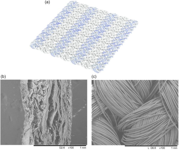
Integrity® consists of approximately 80% chemically modified hyaluronic acid and 20% polyethylene terephthalate (PET). The implant is intended to provide a temporary matrix for fibroblast infiltration and collagen deposition while gradually degrading over several months.
The current clinical evidence cited in the presentation is largely based on manufacturer-provided data rather than robust independent clinical studies.
What Does the Evidence Show for Synthetic Augmentation?
The clinical evidence is strongest for REGENETEN®, particularly in partial-thickness tears.
- In a 2016 study by Bokor et al. [3], 13 patients with intermediate- and high-grade partial-thickness tears underwent isolated implantation of regenten. At 24 months, Constant and ASES scores improved significantly, with increased tendon thickness at early follow-up, although slight decrease in thickness was noted at 24 months. Seven of 13 patients demonstrated complete healing by 12 months, while three demonstrated partial healing.
Dai et al. [4] subsequently reported 24 patients with partial-thickness tears involving more than 50% of tendon thickness. ASES scores increased from 45.6 to 68.1, while VAS scores decreased from 8.3 to 3.8. Tendon thickness also increased from 5.7 mm preoperatively to 6.5 mm at mean follow‐up of 9.9 months.
Schlegel et al. [5] in a study of 33 patients treated with isolated implantation of Regeneten® membrane with 12 patients intermediate‐grade (25%–50%) tears, and 21 patients high grade (>50%) tears reported MRI complete healing in only 18% of cases.
Doyle et al. [6] retrospectively reviewed subjects with high‐grade (Ellmann 3) partial‐thickness posterosuperior tear with at least 12 months follow‐up comparing isolated Regeneten® implantation with completion of tear and repair. They did not find relevant clinical differences at 12 months follow‐up with a rate of revision repair at final follow‐up 2.1% versus 5.4%.
However, the evidence should be interpreted cautiously. Much of the literature consists of case series and retrospective studies, and therefore does not provide the same level of evidence as well-designed randomized controlled trials.
Synthetic Augmentation in Full-Thickness Tears: Not a Uniform Success Story
The results become more heterogeneous when synthetic augmentation is used with repaired full-thickness tears.
Bokor et al. [7] reported substantial improvements in Constant (from 50.7 to 78; p < 0.001) and ASES (from 44.6 to 87.8; p < 0.001) scores following repair augmented with REGENETEN®, together with increased tendon thickness. slight reduction of tendon thickness was observed at 24 months
Bushnell et al. [8] reported MRI healing in 95% of medium-sized tears and 65% of larger tears at two years.
However, randomized and comparative data have not consistently demonstrated a clinical advantage.
In a two-arm randomized study involving 124 patients with small-to-medium tears, Ruiz Ibán et al. [9] found no significant difference between repair with and without REGENETEN® augmentation in achieving the MCID for ASES and Constant-Murley scores.
Similarly, a retrospective matched [10] study reported no significant clinical difference at two years but found a higher reoperation rate in the augmented group, primarily related to stiffness and inflammation causing increased reoperation rate (9% vs. 0%, p = 0.01)
For large and massive tears, the evidence is also inconclusive. One comparative study by Zhang et al. [11] reported healing rates of 55% with REGENETEN® versus 53% without augmentation, with no significant difference in clinical or MRI outcomes.
These findings highlight an important principle:
Increasing tendon thickness does not necessarily translate into improved functional outcomes or lower re-tear rates.
The Other Side of the Equation: Complications
Although modern synthetic implants are designed to be biocompatible and resorbable, foreign-body-related complications remain relevant.
Reported complications include:
- Postoperative stiffness
- Inflammatory reactions
- Foreign-body reaction
- Subacromial inflammation
- Implant migration
- Need for subsequent debridement
In one retrospective study by Yeazell et al. [12], postoperative stiffness at three months occurred in 25% of patients receiving REGENETEN® compared with 3% of controls, and 18% required additional intervention for stiffness.
Ting et al. [13] compared outcomes in 19 patients who underwent RCT with Regeneten® and 32 patients without augmentation. At 6‐week follow‐up, control group showed greater external rotation and forward flexion. Similarly, at 3‐month follow‐up, greater forward flexion observed in nonaugmented group. At final follow‐up, retear rate in Regeneten® group 47%, not significantly different from control, with retear rate of 38% .
Severe inflammatory reactions with rice-body formation [14] have also been reported, although cumulative incidence of this complication in larger studies appears to be less than 1%.
Cost is another consideration. approximate implant costs of:
| Implant | Approximate cost |
| REGENETEN® | $3,000 |
| BioBrace® | $2,000 |
| Tapestry® | $2,000–3,500 |
| Integrity® | $3,000–5,000 |
This creates an important practical question: Can comparable or superior biological augmentation be achieved using tissue already available during surgery?
Autograft Augmentation: Using Biology Instead of Manufacturing It
The most interesting alternative is the use of autologous tissue, particularly the long head of the biceps tendon.
The LHBT is readily accessible through the same surgical field, has a vascularized cellular component and can be incorporated into the reconstruction without the need for a separate donor site.
Its potential mechanisms include:
- Mechanical reinforcement
- Reduction of tension at the tendon-bone interface
- Biological contribution from tenocytes and fibroblasts
- Additional collagen production
- Potential contribution to vascularization and remodeling
- Mechanical bumper effect
- Management of concomitant biceps pathology
This is particularly attractive because the LHBT can simultaneously address a common source of anterior shoulder pathology while contributing tissue to the cuff reconstruction.
Long Head of Biceps Tendon Augmentation
First to use of the LHBT as an augmentation in rotator cuff reconstruction was Dr. Neviaser in 1971. Subsequent techniques have used the intra-articular portion of the LHBT after tenotomy as a biological augmentation for massive rotator cuff reconstruction.
Rhee et al. [15] reported on 16 patients undergoing this approach, with a mean follow-up of 32 months. The average Constant-Murley score was 83.5, and MRI demonstrated healing in approximately 64.3% of patients.
More recent comparative studies have suggested potentially favourable structural outcomes when LHBT augmentation is incorporated into repair.
One study [16] reported re-tear rate of partial repair + LHBT augmentation 43.3% vs partial repair alone 73.3% favoring augmentation
Another comparison of rotator cuff repair with superior capsular reconstruction versus repair with LHBT tenotomy [17] reported re-tear rates of 16.7% versus 40.9%, respectively.
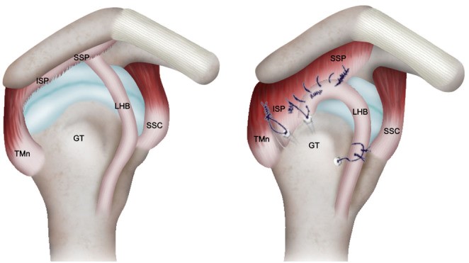
A further study [18] reported complete healing in 58.3% of patients receiving LHBT augmentation compared with 26.3% without augmentation.
These results are encouraging, although the heterogeneity of surgical techniques and study designs means that they should not be interpreted as definitive proof of superiority.
Why LHBT Augmentation Is Attractive
The practical advantages of LHBT augmentation are particularly compelling.
It is immediately available
The graft is obtained from the operative field and does not require harvesting from a separate anatomical site.
It is autologous
There is no cadaveric tissue and no synthetic foreign-body scaffold.
It requires minimal preparation
The tendon can be incorporated into the repair without complex graft preparation.
It may simultaneously address biceps pathology
Tenodesis following use of the LHBT can treat associated biceps disease while contributing to the cuff reconstruction.
It is cost-effective
Unlike commercially manufactured implants, the tissue itself does not carry an additional implant cost.
The presentation summarizes these advantages as:
FREE – NON-CADAVERIC – NO PREPARATION NEEDED – DOES NOT PREVENT BICEPS TENODESIS.
Clinical Outcomes With LHBT Augmentation
The clinical results presented are encouraging.
One series [19] involving 22 patients evaluated by MRI/USG reported a 4% re-tear rate at an average follow-up of 23.5 months, with a mean ASES score of 96.
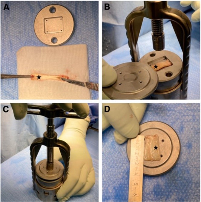
Another study [20] of 46 patients reported re-rupture in only three patients (6.5%). Constant-Murley scores improved from 40.30 to 79.54, ASES scores from 23.89 to 77.48, and VAS scores from 7.37 to 2.76.
In a larger comparative cohort [21] of 77 patients, the reported one-year re-rupture rates were 38.5% in the control group vs 16% with LHBT augmentation
While these findings are promising, they should be interpreted in the context of study design, patient selection and variation in augmentation techniques.
Fascia Lata: Another Autologous Option
The concept of autologous augmentation extends beyond the biceps.
Fascia lata has been used as a biological graft in large-to-massive rotator cuff tears, particularly in patients with advanced fatty degeneration or recurrent tears.
A series of 14 patients [22] with large-to-massive tears and Goutallier grade 3–4 fatty degeneration or retears with one year follow up reported:
- VAS improved from 5.1 to 1.3
- ASES improved from 51.9 to 85.2
- Constant score improved from 51.3 to 70.8
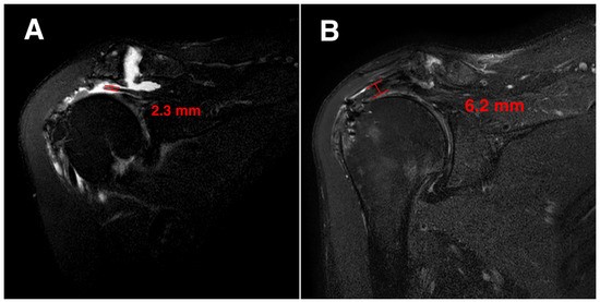
These improvements were maintained through five years, with a reported re-tear rate of 7.1% (1/14) and no revision surgery during follow-up.
MRI evidence was also noted of substantial postoperative tendon thickening in a patient with a massive cuff tear, from 2.3 mm preoperatively to 6.2 mm at 12 months.
Autograft Versus Synthetic Augmentation:
Both approaches attempt to solve different problems.
| Feature | Synthetic/Bioinductive Implant | Autograft |
| Tissue source | Manufactured/animal-derived scaffold | Patient’s own tissue |
| Biological potential | Depends on scaffold and host response | Living autologous tissue |
| Mechanical reinforcement | Variable; implant dependent | Variable |
| Foreign-body exposure | Present | Absent |
| Preparation | Implant-specific | Usually minimal |
| Donor-site morbidity | None | Depends on graft source |
| Cost | Often substantial | Minimal additional graft cost |
| Biceps pathology | Does not directly address it | LHBT augmentation can address it |
| Evidence base | Strongest for REGENETEN®, but heterogeneous | Increasing but still heterogeneous |
| Long-term evidence | Variable | Still evolving |
| Main limitation | Cost and implant-related reactions | Limited high-level comparative evidence |
The critical issue is therefore not whether autograft augmentation should replace every synthetic implant.
Rather, it is whether a readily available autologous tendon can provide sufficient biological and mechanical augmentation in selected patients without the cost and potential complications associated with manufactured scaffolds.
Is Autograft Actually “Better”?
The evidence presented in the current literature is promising but does not yet establish universal superiority of autograft augmentation over synthetic augmentation.
Synthetic implants, particularly REGENETEN®, have a substantially larger published clinical experience than some of the newer scaffolds. However, comparative studies have not consistently demonstrated improved functional outcomes or reduced re-tear rates with synthetic augmentation.
Autograft techniques offer several theoretical and practical advantages:
- Autologous living tissue
- No manufactured foreign-body scaffold
- Minimal additional cost
- No separate graft-harvesting site when LHBT is used
- Potential biological contribution to healing
- Ability to address concomitant biceps pathology
- Potential mechanical reinforcement
At the same time, the current evidence for autograft augmentation is also dominated by case series and retrospective comparative studies, and techniques vary considerably.
Therefore, the appropriate conclusion is not that every rotator cuff repair should be augmented with autograft.
Instead:
When augmentation is indicated, autologous tissue—particularly the LHBT—represents a biologically attractive, readily available and potentially cost-effective option that deserves serious consideration alongside synthetic implants.
The Future: From Scaffolds to Living Augmentation
The evolution of rotator cuff augmentation is moving from purely mechanical reinforcement toward biological restoration.
Synthetic implants attempt to create an environment in which host cells migrate, proliferate and generate new tendon-like tissue. Autografts begin with the patient’s own living tissue and therefore offer an intrinsically biological substrate.
This distinction may become increasingly important as our understanding of tendon healing improves.
The future may not involve choosing a single “best” augmentation material. Instead, augmentation may become patient-specific, based on:
Tendon quality + tear size + tissue biology + mechanical demands + biceps status + available autologous tissue + cost.
Take-Home Message
Synthetic augmentation has introduced an important new dimension to rotator cuff surgery, with REGENETEN® currently having the most substantial clinical evidence among the newer implants. However, clinical studies have produced mixed results, particularly when synthetic augmentation is added to repair of full-thickness and large/massive tears.
Autologous augmentation, especially using the long head of the biceps tendon, offers a compelling alternative. It is readily accessible, biologically active, inexpensive and can simultaneously address associated biceps pathology.
The available evidence suggests that LHBT augmentation may improve structural healing and reduce re-tear rates in selected high-risk repairs, but higher-quality prospective randomized comparative studies are required before declaring autograft augmentation superior to synthetic augmentation.
For informative videos related to Shoulder problems and their treatment options, Sports Injuries and other orthopedic conditions, visit our YouTube channel Bangalore Shoulder Institute – https://www.youtube.com/@BangaloreShoulderInstitute
REFERENCES
- Graham P. Rotator Cuff Tear. Orthop Nurs. 2018 Mar/Apr;37(2):154-156. doi: 10.1097/NOR.0000000000000441. PMID: 29570550.
- Minagawa H, Yamamoto N, Abe H, Fukuda M, Seki N, Kikuchi K, Kijima H, Itoi E. Prevalence of symptomatic and asymptomatic rotator cuff tears in the general population: From mass-screening in one village. J Orthop. 2013 Feb 26;10(1):8-12. doi: 10.1016/j.jor.2013.01.008. PMID: 24403741; PMCID: PMC3768248.
- Bokor DJ, Sonnabend D, Deady L, Cass B, Young A, Van Kampen C, Arnoczky S. Evidence of healing of partial-thickness rotator cuff tears following arthroscopic augmentation with a collagen implant: a 2-year MRI follow-up. Muscles Ligaments Tendons J. 2016 May 19;6(1):16-25. doi: 10.11138/mltj/2016.6.1.016. PMID: 27331028; PMCID: PMC4915456.
- Dai A, Campbell A, Bloom D, Baron S, Begly J, Meislin R. Collagen-Based Bioinductive Implant for Treatment of Partial Thickness Rotator Cuff Tears. Bull Hosp Jt Dis (2013). 2020 Sep;78(3):195-201. PMID: 32857027.
- Schlegel TF, Abrams JS, Angelo RL, Getelman MH, Ho CP, Bushnell BD. Isolated bioinductive repair of partial-thickness rotator cuff tears using a resorbable bovine collagen implant: two-year radiologic and clinical outcomes from a prospective multicenter study. J Shoulder Elbow Surg. 2021 Aug;30(8):1938-1948. doi: 10.1016/j.jse.2020.10.022. Epub 2020 Nov 19. PMID: 33220413.
- Doyle TR, Hurley ET, Lorentz SG, Briggs DV, Cullen M, Klifto CS, Anakwenze O. Clinical Outcomes of Arthroscopic Treatment of High-Grade Partial Thickness Rotator Cuff Tears With Augmentation Using Bioinductive Collagen Implants Are Comparable With Tear Completion and Repair. Arthroscopy. 2025 Oct;41(10):3918-3926. doi: 10.1016/j.arthro.2025.03.043. Epub 2025 Apr 29. PMID: 40311949.
- Bokor DJ, Sonnabend D, Deady L, Cass B, Young A, Van Kampen C, Arnoczky S. Evidence of healing of partial-thickness rotator cuff tears following arthroscopic augmentation with a collagen implant: a 2-year MRI follow-up. Muscles Ligaments Tendons J. 2016 May 19;6(1):16-25. doi: 10.11138/mltj/2016.6.1.016. PMID: 27331028; PMCID: PMC4915456.
- Bushnell BD, Connor PM, Harris HW, Ho CP, Trenhaile SW, Abrams JS. Retear rates and clinical outcomes at 1 year after repair of full-thickness rotator cuff tears augmented with a bioinductive collagen implant: a prospective multicenter study. JSES Int. 2020 Dec 15;5(2):228-237. doi: 10.1016/j.jseint.2020.10.020. PMID: 33681842; PMCID: PMC7910780.
- Ruiz Ibán MA, García Navlet M, Moros Marco S, Diaz Heredia J, Hernando A, Ruiz Díaz R, Vaquero Comino C, Alvarez Villar S, Avila Lafuente JL. Augmentation of a Posterosuperior Cuff Repair With a Bovine Bioinductive Collagen Implant Shows a Lower Retear Rate but Similar Outcomes Compared With No Augmentation: 2-Year Results of a Randomized Controlled Trial. Arthroscopy. 2025 Oct;41(10):3869-3879. doi: 10.1016/j.arthro.2025.03.057. Epub 2025 Apr 8. PMID: 40209829.
- Haft M, Li SS, Pearson ZC, Ahiarakwe U, Bettencourt AF, Srikumaran U. No Short-term Clinical Benefit to Bovine Collagen Implant Augmentation in Primary Rotator Cuff Repair: A Matched Retrospective Study. Clin Orthop Relat Res. 2025 Mar 1;483(3):442-452. doi: 10.1097/CORR.0000000000003247. Epub 2024 Sep 5. PMID: 39236102; PMCID: PMC11828003.
- Zhang T, Ajayi A, Hajjar M, Fleckenstein CM, Nolan J, Hasan SS. Arthroscopic Repair of Retracted Large and Massive Rotator Cuff Tears With and Without Augmentation With a Bio-Inductive Collagen Implant Reveals Substantial and Comparable Clinical Improvement. Arthroscopy. 2024 May;40(5):1434-1442. doi: 10.1016/j.arthro.2023.10.024. Epub 2023 Nov 4. PMID: 37931669.
- Yeazell S, Lutz A, Bohon H, Shanley E, Thigpen CA, Kissenberth MJ, Pill SG. Increased stiffness and reoperation rate in partial rotator cuff repairs treated with a bovine patch: a propensity-matched trial. J Shoulder Elbow Surg. 2022 Jun;31(6S):S131-S135. doi: 10.1016/j.jse.2022.02.003. Epub 2022 Mar 12. PMID: 35288296.
- Iban MÁR, Reina MJE, Del Caño CD, Villar SÁ, Heredia JD, Lafuente JLÁ. The role of new synthetic membranes in rotator cuff augmentation. J Exp Orthop. 2026 Jan 21;13(1):e70634. doi: 10.1002/jeo2.70634. PMID: 41574209; PMCID: PMC12820577.
- Barad SJ. Severe subacromial-subdeltoid inflammation with rice bodies associated with implantation of a bio-inductive collagen scaffold after rotator cuff repair. J Shoulder Elbow Surg. 2019 Jun;28(6):e190-e192. doi: 10.1016/j.jse.2019.02.019. Epub 2019 Apr 16. PMID: 31003886.
- Rhee YG, Cho NS, Lim CT, Yi JW, Vishvanathan T. Bridging the gap in immobile massive rotator cuff tears: augmentation using the tenotomized biceps. Am J Sports Med. 2008 Aug;36(8):1511-8. doi: 10.1177/0363546508316020. Epub 2008 Apr 28. PMID: 18443279.
- Laprus H, Brzóska R, Błasiak A, Juszczak B, Malik SS, Lubiatowski P. Massive and irreparable rotator cuff tear treatment by arthroscopic partial repair with long head of the biceps tendon augmentation provides better healing and functional results than partial repair only. J Shoulder Elbow Surg. 2024 Feb;33(2):263-272. doi: 10.1016/j.jse.2023.06.022. Epub 2023 Jul 22. PMID: 37482246.
- Chiang CH, Shaw L, Chih WH, Yeh ML, Ting HH, Lin CH, Chen CP, Su WR. Modified Superior Capsule Reconstruction Using the Long Head of the Biceps Tendon as Reinforcement to Rotator Cuff Repair Lowers Retear Rate in Large to Massive Reparable Rotator Cuff Tears. Arthroscopy. 2021 Aug;37(8):2420-2431. doi: 10.1016/j.arthro.2021.04.003. Epub 2021 Apr 15. PMID: 33864834.
- Cho NS, Yi JW, Rhee YG. Arthroscopic biceps augmentation for avoiding undue tension in repair of massive rotator cuff tears. Arthroscopy. 2009 Feb;25(2):183-91. doi: 10.1016/j.arthro.2008.09.012. Epub 2008 Nov 1. PMID: 19171279.
- Brinkman JC, Moore ML, Smith C, Tummala SV, Tokish JM. Rotator Cuff Repair Augmentation With an Autograft Biceps Patch, the “Biceps Smash,” Results in Low Retear Rates at 6 Months and Promising Clinical Outcomes at 12 Months. Arthroscopy. 2026 Aug;42(8):1347-1354. doi: 10.1002/arj.70379. Epub 2026 Jun 15. PMID: 42295852.
- Karakus O, Oztermeli A. Clinical outcomes of long head of the biceps tendon augmentation versus partial repair in massive rotator cuff tears. J Orthop Surg Res. 2026 May 22;21(1):509. doi: 10.1186/s13018-026-06959-1. PMID: 42174664; PMCID: PMC13520536.
- Pastor PCS, Ramos IP, Roig AG, Safont JA. Long head of biceps tendon augmentation in rotator cuff repair enhances tendon healing, shoulder function and patient-reported outcomes one-year post-surgery. J Exp Orthop. 2024 Oct 10;11(4):e70033. doi: 10.1002/jeo2.70033. PMID: 39391567; PMCID: PMC11465290.