Written by: Editorial Team of Dr Ayyappan V Nair
Medically Reviewed by: Dr. Ayyappan V. Nair (https://drayyappanvnair.com)
Senior Consultant – Shoulder Surgery, Arthroscopy and Sports Trauma
Manipal Hospitals, Bangalore
Last Reviewed: 8th August 2026
Calcific shoulder tendonitis happens when calcium crystals build up inside a rotator cuff tendon, most often the supraspinatus, without any injury triggering it. It moves through distinct biological stages, and the worst pain usually strikes not while the deposit is forming but when the body starts breaking it down. Diagnosis relies on X-ray and ultrasound rather than MRI alone. Treatment ranges from rest and physiotherapy to needle aspiration (barbotage) and, in a smaller group of patients, arthroscopic removal. Knowing which stage a patient is in changes the entire management plan, which is exactly why this condition gets mismanaged so often.
A patient walks in describing shoulder pain that came on almost overnight, sometimes waking them from sleep, with no fall, no lifting incident and no obvious trigger at all. This catches most people off guard, since nearly every other shoulder problem they have heard of starts with an injury. Calcific tendonitis does not work that way, and understanding why changes how it should be treated.
What Is Actually Happening Inside the Tendon
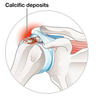
Calcific tendonitis develops when calcium hydroxyapatite crystals deposit inside the fibres of a rotator cuff tendon, most commonly the supraspinatus, the tendon running across the top of the shoulder. This is not the same process as the calcium deposits seen in arthritis or in a chronic tendon tear. It is a distinct, self-contained biological event that follows its own timeline, largely independent of how much the shoulder has been used.
The exact trigger is not fully understood. One theory is that changes within the tendon cause some cells to behave more like cartilage cells, creating conditions in which calcium hydroxyapatite can collect. Once that deposit is established, the tendon’s normal gliding mechanism under the acromion is disrupted, and this is where the mechanical irritation and pain begin.
The Three Stages, and Why the Pain Pattern Flips
This condition moves through three recognised phases, and the relationship between deposit size and pain is counterintuitive enough that it trips up patients and, occasionally, clinicians too.
Stage | What is happening | Typical pain pattern | Deposit appearance |
Formative | Calcium is actively being laid down within the tendon | Dull ache, gradually worsening, often mistaken for early impingement | Dense, well defined, chalky on imaging |
Resting | The deposit is stable and inactive | Often minimal pain, sometimes discovered incidentally on an X-ray for another reason | Unchanged, well demarcated |
Resorptive | The body actively breaks the deposit down and clears it | Sudden, severe, sometimes disabling pain, frequently at night | Fluffy, poorly defined edges, softer consistency |
The mechanism behind that resorptive spike matters clinically. As the body dissolves the deposit, it triggers an intense local inflammatory response, and pressure builds within the tendon as the calcium turns from a solid, toothpaste like consistency into a more liquid one before being cleared. The severe pain during this phase is thought to come from the inflammatory response, rising pressure within the tendon and irritation of the nearby bursa as the deposit softens and breaks down. .
Calcific Tendonitis or a Rotator Cuff Tear?
Patients, and sometimes referring doctors, often assume severe shoulder pain with weakness must mean a torn rotator cuff. Separating these two pathways properly changes the entire treatment approach.
Pathway one: calcific tendonitis with an intact tendon. The tendon fibres remain structurally continuous. Pain and weakness come from inflammation, swelling and mechanical irritation around the calcium deposit, not from an actual structural gap in the tendon. Strength typically improves quickly once the acute inflammatory phase settles, even before the deposit fully clears.
Pathway two: a genuine rotator cuff tear. Here, tendon fibres are physically disrupted, whether partial or full thickness. Weakness tends to be more persistent and does not resolve simply with pain control, because the mechanical pulley system of the shoulder is actually compromised, not just irritated.
The consequence of mixing these two up is significant. Recommending early surgical repair for what is actually calcific tendonitis exposes a patient to an unnecessary operation, when many of these cases settle with far simpler treatment. Conversely, dismissing a genuine tear as calcific inflammation delays repair and can allow the tear to progress. An ultrasound or MRI, read alongside a plain X-ray, is what reliably tells the two apart, since calcific deposits show up clearly on X-ray in a way that a straightforward tear does not.
How Doctors Confirm It’s Calcific Tendonitis
A shoulder X-ray remains the most reliable first step, since it shows the deposit’s size, density and location clearly, and gives a strong clue as to which stage the deposit is in based on how well defined its edges appear. Ultrasound adds real time information about deposit consistency and can guide treatment directly during the same appointment. MRI is generally reserved for cases where a coexisting rotator cuff tear or other pathology needs ruling out, rather than as a routine first test.
Treatment Options, Matched to the Stage
- Formative or resting stage with manageable pain: anti-inflammatory medication, activity modification and a structured physiotherapy programme focused on maintaining range of motion, since aggressive treatment at this stage often is not necessary.
- Resorptive stage with severe pain: pain relief and inflammation control are usually the immediate priorities. Ultrasound-guided barbotage may be considered when symptoms remain severe, depending on the deposit’s consistency and the specialist’s assessment.
- Persistent symptoms despite non-surgical treatment: extracorporeal shockwave therapy is used in some cases to help fragment a stubborn deposit and stimulate healing, particularly useful for deposits that have settled into a stable but symptomatic resting phase.
- Deposits resistant to all conservative measures: arthroscopic removal is reserved for a genuinely small subset of patients, typically those with longstanding symptoms that have not responded to repeated non-surgical attempts over several months.
Most patients never need to reach the final step on that list. The natural history of this condition, particularly once it enters the resorptive phase, tends towards genuine resolution rather than a chronic, worsening problem, which is precisely why treatment intensity should be matched carefully to the stage rather than jumping straight to the most aggressive option out of frustration with the pain.
Making Sense of Sudden, Unexplained Shoulder Pain
Severe shoulder pain with no clear cause is unsettling, and it is entirely reasonable to assume the worst. A shoulder X-ray can often identify the deposit and help guide the next steps, although the findings still need to be considered alongside the patient’s symptoms and examination.
FAQs
Does calcific tendonitis mean I will eventually need surgery?
No. The large majority of cases resolve with non-surgical treatment, particularly once the deposit reaches the resorptive stage and the body clears it naturally. Surgery is reserved for a small minority with persistent symptoms despite proper conservative management.
Why is the pain worse at night?
Night pain is common during the inflammatory phase, particularly when lying on the affected side, because pressure on the irritated tendon and bursa can aggravate symptoms
Can calcific tendonitis come back after treatment?
It can occur again in the same shoulder or the opposite one, since the underlying cause relates to tendon biology rather than a mechanical injury that has been fixed. However, a fully resolved and cleared deposit does not typically return in the same location.
For enquiries and online appointments, send a message to www.DrAyyappanVNair.com/contact
For informative videos related to Shoulder problems and their treatment options, Sports Injuries and other orthopedic conditions, visit our YouTube channel Bangalore Shoulder Institute – https://www.youtube.com/@BangaloreShoulderInstitute