Written by: Editorial Team of Dr Ayyappan V Nair
Medically Reviewed by: Dr. Ayyappan V. Nair
Senior Consultant – Shoulder Surgery, Arthroscopy and Sports Trauma
Manipal Hospitals, Bangalore
Last Reviewed: 12th September 2026
Introduction
The biceps brachii is a powerful muscle of the arm that plays an important role in elbow flexion and forearm supination, while also contributing to shoulder function.
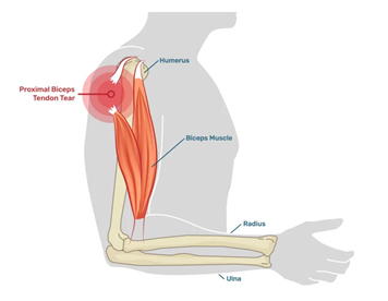
At the shoulder, the biceps has two proximal heads:
- Long head of the biceps (LHB) — originates from the superior glenoid/labral complex and travels through the bicipital groove.
- Short head of the biceps — originates from the coracoid process.
Distally, the two muscle bellies unite and form a common distal tendon that inserts primarily on the radial tuberosity, with an additional aponeurotic expansion through the lacertus fibrosus.
A biceps tendon rupture occurs when the tendon partially or completely fails. Depending on the anatomical location, the injury is classified as:
Proximal biceps tendon rupture
Usually involves the long head of the biceps at the shoulder.
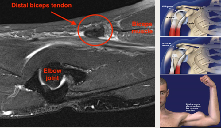
Distal biceps tendon rupture
Occurs at the elbow, where the distal tendon detaches from the radial tuberosity.
Although both are termed “biceps tendon ruptures,” they are not equivalent injuries.
A proximal long-head rupture is frequently tolerated functionally, particularly in older, lower-demand individuals. In contrast, an acute complete distal biceps rupture can produce clinically important loss of supination and elbow-flexion strength, and surgical repair is commonly considered in active patients, says Dr Ayyappan V Nair, considered as the best shoulder surgeon in Bangalore.
Proximal Versus Distal Biceps Rupture
Understanding this distinction is fundamental to treatment planning.
Feature | Proximal long-head rupture | Distal biceps rupture |
Typical location | Shoulder | Elbow |
Common patient profile | Older patients; degenerative tendon disease | Active, often middle-aged men |
Mechanism | Often degenerative or relatively low-energy | Usually sudden eccentric load |
Visible deformity | Popeye sign | Proximal retraction/bulge |
Main functional deficit | Usually modest | Significant supination and flexion weakness |
Initial treatment | Often non-operative | Surgery frequently considered in active patients |
Common surgery | Tenodesis in selected patients | Anatomic tendon reinsertion |
This distinction also explains why the statement that “biceps tendon rupture should always be operated on early” is incorrect.
Causes and Risk Factors
Biceps tendon rupture may result from either degenerative tendon disease or acute trauma.
Proximal rupture
The long head of the biceps is exposed to repetitive mechanical loading as it passes through the shoulder joint and bicipital groove.
Risk factors include:
- Increasing age
- Rotator cuff disease
- Chronic biceps tendinopathy
- Repetitive overhead activity
- Heavy lifting
- Smoking
- Previous shoulder pathology
- Repetitive sports activity
Proximal rupture often represents the final stage of chronic tendon degeneration rather than an isolated acute injury.
Distal rupture
Distal biceps rupture typically occurs during a sudden eccentric contraction, for example when attempting to control a heavy load while the elbow is forced toward extension.
Common scenarios include:
- Weight lifting
- Sudden heavy lifting
- Sports
- Occupational injuries
- High-load eccentric activity
The classic patient is an active middle-aged man who experiences a sudden pain or “pop” at the elbow while lifting.
Clinical Presentation
Proximal Long-Head Rupture
Patients may describe:
- Sudden anterior shoulder or upper-arm pain
- A snapping or popping sensation
- Bruising
- Weakness or fatigue with lifting
- Cramping discomfort
- Visible change in the contour of the upper arm
The classic finding is the Popeye deformity, produced by distal retraction of the long-head muscle belly.
However, the absence of a dramatic Popeye sign does not exclude a proximal rupture.
Patients may also have coexisting:
- Rotator cuff tears
- Subacromial pathology
- Glenohumeral arthritis
- Long-head biceps tendinopathy
Therefore, the biceps should not be assessed in isolation.
Distal Biceps Rupture
The presentation is often much more dramatic.
Typical findings include:
- Sudden pain in the antecubital fossa
- Audible or palpable “pop”
- Bruising around the elbow and proximal forearm
- Weakness of elbow flexion
- Marked weakness of forearm supination
- Visible proximal retraction of the biceps muscle
- Loss of the normal distal biceps contour
The loss of supination strength is particularly important.
Activities such as:
- Turning a screwdriver
- Opening a tight jar
- Using a wrench
- Carrying a heavy object with the palm facing upward
may become difficult.
Diagnosis
Diagnosis begins with a careful history and physical examination.
Clinical Examination
For proximal pathology, examination may reveal tenderness over the bicipital groove, pain with resisted elbow flexion or forearm supination, and a Popeye deformity.
For distal rupture, specific examination manoeuvres are useful.
Hook Test
The elbow is flexed and the examiner attempts to “hook” a finger underneath the distal biceps tendon from the lateral side.
A normally positioned tendon can usually be palpated.
An absent or abnormal hook test raises strong suspicion of distal biceps rupture.
Biceps Squeeze Test
Compression of the biceps muscle belly normally produces forearm supination.
Failure to produce the expected response suggests distal tendon disruption.
No single clinical test should be interpreted in isolation, particularly in partial tears.
Imaging
X-rays
Plain radiographs do not directly demonstrate the biceps tendon.
However, they can help identify:
- Avulsion-related bony changes
- Radial tuberosity pathology
- Associated fractures
- Other causes of elbow or shoulder pain
Ultrasound
High-resolution ultrasound can be particularly useful for evaluating the biceps tendon dynamically.
It can demonstrate:
- Tendon discontinuity
- Retraction
- Fluid surrounding the tendon
- Partial tears
- Long-head biceps pathology
- Associated rotator cuff abnormalities
Ultrasound also provides a rapid, relatively inexpensive dynamic assessment.
MRI
MRI is particularly useful when:
- The diagnosis is uncertain
- A partial tear is suspected
- The extent of tendon retraction needs assessment
- Associated shoulder or elbow pathology must be evaluated
- Surgical planning is required
MRI can also help differentiate a complete rupture from severe tendinopathy.
Treatment
Treatment should be based on the anatomical location of the rupture, patient age, activity level, functional requirements, associated pathology and expectations.
The treatment strategy for a proximal rupture is very different from that for a distal rupture.
Proximal Long-Head Biceps Rupture
Non-operative Treatment
For many patients, particularly older or lower-demand individuals, treatment can be conservative.
Options include:
- Activity modification
- Analgesics/NSAIDs when appropriate
- Physiotherapy
- Restoration of shoulder range of motion
- Rotator cuff and scapular strengthening
- Management of associated shoulder pathology
A proximal long-head rupture often does not produce a major functional deficit because the short head remains intact and the biceps retains substantial function.
Therefore, the presence of a Popeye deformity alone is generally not an indication for surgery.
When Is Surgery Considered for Proximal Biceps Rupture?
Surgery may be considered in selected patients with:
- Persistent pain attributable to the biceps
- Significant cramping
- Symptomatic instability
- High functional demands
- Cosmetic concerns
- Failure of appropriate non-operative treatment
- Associated rotator cuff or labral pathology requiring surgery
The two principal surgical options are:
Tenotomy
The diseased long head is released and allowed to retract.
Advantages:
- Technically simple
- Shorter operative time
- No fixation required
Potential disadvantages:
- Popeye deformity
- Possible cramping
- Potential alteration in biceps contour
Tenodesis
The long head of the biceps is released from its origin and fixed to the humerus.
Advantages:
- Reduces the likelihood of Popeye deformity
- Preserves a more normal biceps contour
- May be preferable in younger or physically active patients
Potential disadvantages:
- More technically demanding
- Longer operative time
- Risks associated with fixation
Tenotomy Versus Tenodesis: What Does the Evidence Show?
The debate between tenotomy and tenodesis continues.
A systematic review and meta-analysis of 11 randomized controlled trials involving 860 patients found that overall patient-reported functional outcomes and pain were comparable between the two procedures.
However, tenodesis substantially reduced the rate of Popeye deformity.
“Tenodesis resulted in a lower rate of Popeye deformity.”
— BMJ Open, systematic review and meta-analysis
The same analysis found that tenotomy had a shorter operative time.
Other meta-analyses have similarly found better preservation of certain aspects of biceps function and a lower incidence of Popeye deformity with tenodesis, although differences in overall clinical outcomes are often modest.
Practical message
Tenotomy is not a “wrong” operation and tenodesis is not automatically “better.”
The choice should consider:
- Age
- Activity level
- Cosmetic expectations
- Dominant arm
- Occupation
- Associated rotator cuff pathology
- Risk of cramping
- Surgeon experience
Distal Biceps Tendon Rupture
The management philosophy changes substantially for distal rupture.
A complete distal biceps rupture can result in meaningful loss of:
- Forearm supination strength
- Elbow-flexion strength
- Endurance
This is particularly relevant for manual workers, athletes and physically active patients.
Non-operative Treatment
Non-operative treatment may be appropriate for:
- Low-demand patients
- Older individuals with limited functional requirements
- Patients with significant medical comorbidities
- Selected partial tears
- Patients who decline surgery
However, patients should be counselled that conservative management may result in persistent strength deficits.
A 2024 systematic review of partial distal biceps tears found that conservative treatment was associated with lower complication rates and higher satisfaction, but surgical treatment generally provided better strength outcomes.
Therefore, partial tears require an individualized decision rather than an automatic surgical approach.
Surgical Repair of Complete Distal Biceps Rupture
For an active patient with an acute complete distal biceps rupture, anatomic surgical reinsertion is commonly recommended when restoration of strength is an important goal.
The tendon is retrieved and reattached to the radial tuberosity using fixation techniques such as:
- Cortical button fixation
- Suture anchors
- Bone tunnels
- Other contemporary fixation constructs
The surgical approach may be:
- Single incision
- Two incision
The choice depends on surgeon preference, tendon retraction, anatomy and fixation strategy.
Does Timing Matter?
An acute rupture is generally easier to repair because the tendon has undergone less retraction and scarring.
However, the old statement that every distal rupture must be repaired immediately or it becomes impossible to treat is too absolute.
A systematic review of delayed distal biceps repairs found that when sufficient tendon excursion and tendon quality remain, primary repair can still provide good satisfaction, range of motion and functional outcomes even after delayed presentation.
Nevertheless, chronic tears may require more extensive dissection and may have a greater risk of complications. Some chronic cases may require graft reconstruction when the tendon cannot be mobilized adequately.
Practical message
Early assessment is important even when surgery is not performed immediately.
Modern Evidence on Distal Biceps Repair
A 2025 systematic review evaluating return to sport after distal biceps repair included 42 studies and 1,093 patients.
The overall return-to-sport rate was approximately 91.5%, with approximately 85% returning to their pre-injury level or higher.
The mean time to return to sport was approximately 6.3 months.
“There was a high rate of RTS after DBT repair at 6 months postoperatively.”
— Boufadel et al., American Journal of Sports Medicine, 2025
This provides useful information when counselling athletic patients.
Rehabilitation After Distal Biceps Repair
Rehabilitation has traditionally involved a period of protected immobilization followed by progressive restoration of movement and strength.
The rehabilitation programme generally progresses through:
Phase 1 — Protection
- Splint or hinged brace
- Protection of the repair
- Controlled range of motion
- Avoidance of resisted elbow flexion and supination
Phase 2 — Restoration of Motion
- Progressive elbow range of motion
- Gradual forearm rotation
- Reduction in protective restrictions according to healing
Phase 3 — Strengthening
- Progressive biceps loading
- Forearm strengthening
- Functional strengthening
- Gradual return to occupational or sporting activities
Phase 4 — Return to Sport/Heavy Activity
Strength and endurance are progressively restored before unrestricted return to high-load activities.
Is Early Mobilization Safe?
This is an evolving area of rehabilitation research.
A 2025 systematic review and meta-analysis examining postoperative rehabilitation after distal biceps repair found no clinically significant difference in patient-reported outcomes or range of motion between early and delayed mobilization protocols.
A separate 2026 systematic review of 50 studies involving 1,577 patients similarly found no significant difference in re-rupture rates, DASH scores or strength ratios between early mobilization and immobilization groups, although the certainty of evidence was low to very low.
Clinical implication
The evidence does not currently support one universal rehabilitation protocol.
Instead, rehabilitation should be:
Repair-specific + fixation-specific + patient-specific.
The biological healing of the tendon-bone interface must remain the central consideration.
Complications of Distal Biceps Surgery
Patients should be counselled regarding potential complications, including:
- Nerve irritation or neuropraxia
- Lateral antebrachial cutaneous nerve symptoms
- Posterior interosseous nerve injury
- Heterotopic ossification
- Radioulnar synostosis
- Loss of forearm rotation
- Infection
- Re-rupture
- Persistent weakness
- Elbow stiffness
The reported complication profile varies according to surgical approach and fixation technique.
Therefore, surgical counselling should include not only the potential benefits but also the specific risks associated with the chosen approach.
A Practical Treatment Algorithm
Proximal Long-Head Rupture
Clinical assessment + ultrasound/MRI when indicated
↓
Mild symptoms / low-demand patient
Observation + analgesia + physiotherapy
↓
Persistent symptomatic biceps pathology
Consider:
Tenotomy or tenodesis
↓
Associated rotator cuff/labral pathology
Address biceps pathology in conjunction with treatment of the associated lesion
Distal Biceps Rupture
Clinical examination + ultrasound/MRI
↓
Partial tear / low-demand patient
Individualized conservative management
↓
Complete acute rupture in active patient
Discuss early surgical repair
↓
Chronic rupture
Assess:
Tendon excursion + tendon quality + degree of retraction
↓
Primary repair if adequate
or
Reconstruction with graft when primary repair is not feasible
↓
Rehabilitation
Protected early phase → progressive ROM → strengthening → functional rehabilitation → return to sport/work
Evidence Boxes
Evidence Box 1 — Tenotomy vs Tenodesis
11 randomized controlled trials | 860 patients
Overall pain and patient-reported functional outcomes were broadly comparable between tenotomy and tenodesis.
However, tenodesis substantially reduced the risk of Popeye deformity.
Clinical message: Choose the procedure according to patient characteristics and expectations rather than assuming that one operation is universally superior.
Evidence Box 2 — Return to Sport After Distal Repair
42 studies | 1,093 patients
Approximately 91.5% returned to sport, with approximately 85% returning to pre-injury level or higher.
Mean return-to-sport time was approximately 6.3 months.
Clinical message: Surgical repair can provide a high likelihood of return to sport in appropriately selected patients.
Evidence Box 3 — Chronic Distal Ruptures
Delayed repair does not automatically mean reconstruction is necessary.
When sufficient tendon excursion and quality remain, direct repair can still produce good functional outcomes.
Clinical message: Refer and assess early, but do not consider a delayed presentation an automatic contraindication to primary repair.
Evidence Box 4 — Postoperative Rehabilitation
Recent systematic reviews suggest that early and delayed mobilization after distal biceps repair may produce comparable outcomes, although the quality of evidence remains limited.
Clinical message: Rehabilitation should be individualized rather than dictated by a single universal timeline.
Key Takeaways for Orthopaedic Practice
- Proximal and distal biceps ruptures are different clinical entities and should not be managed identically.
- Proximal long-head rupture is often associated with chronic degenerative tendon disease and can frequently be treated non-operatively.
- A Popeye deformity alone is not an indication for surgery.
- Tenotomy and tenodesis both provide good outcomes for symptomatic long-head biceps pathology.
- Tenodesis substantially reduces the likelihood of a Popeye deformity and may be preferred in younger, active or cosmetically concerned patients.
- Complete distal biceps rupture can produce significant loss of supination and flexion strength.
- Active patients with acute complete distal ruptures should generally be counselled regarding surgical repair.
- Early referral is valuable, but delayed distal repair can still achieve good results when adequate tendon excursion is present.
- Chronic irreparable tears may require graft reconstruction.
- Return to sport after distal biceps repair is generally high, with recent evidence suggesting approximately nine out of ten patients return to sport.
- Modern rehabilitation protocols are increasingly moving toward controlled early mobilization, although there is not yet a single universally accepted protocol.
- The ultimate treatment decision should incorporate patient demands, functional goals, tendon pathology, associated injuries and surgeon expertise.
Conclusion
Biceps tendon rupture represents a spectrum of injuries rather than a single clinical entity.
The most important first step is to establish where the rupture has occurred.
A proximal long-head rupture may be surprisingly well tolerated despite an obvious Popeye deformity, whereas a distal rupture can significantly compromise forearm supination and elbow-flexion strength.
Modern management therefore emphasizes patient-specific decision making rather than a one-size-fits-all surgical approach.
For proximal long-head pathology, conservative management remains appropriate for many patients, while tenotomy and tenodesis provide effective surgical options in selected symptomatic individuals.
For complete distal ruptures in active patients, anatomic repair remains an important treatment option, with contemporary evidence demonstrating high rates of functional recovery and return to sport. At the same time, advances in rehabilitation are challenging the traditional concept that prolonged postoperative immobilization is always necessary.
The modern approach to biceps tendon rupture can therefore be summarized in three principles:
Identify the location.
Understand the functional deficit.
Match the treatment to the patient’s demands.
For enquiries and online appointments, send a message to
www.DrAyyappanVNair.com/contact
For informative videos related to Shoulder problems and their treatment options, Sports Injuries and other orthopedic conditions, visit our YouTube channel Bangalore Shoulder Institute – https://www.youtube.com/@BangaloreShoulderInstitute
Selected Recent References
- Boufadel P, Daher M, Lopez R, et al. Return to Sport After Distal Biceps Tendon Repair: A Systematic Review. Am J Sports Med. 2025;53(7):1769-1778.
- Simpson ER, Jawanda H, Patel I, et al. No Clinically Significant Differences in Patient-Reported Outcomes and Range of Motion Between Early and Delayed Mobilization After Primary Distal Biceps Tendon Repair: A Systematic Review and Meta-analysis. Am J Sports Med. 2025.
- Sconza C, et al. Distal biceps tendon repair: immobilization versus early mobilization. A systematic review and meta-analysis. EFORT Open Rev. 2026.
- Clinical effectiveness of tenotomy versus tenodesis for long head of biceps pathology: a systematic review and meta-analysis. BMJ Open. 2022.
- Hopper HM, Nelson CT, Sandoval LT, et al. Decreased Strength, Complication Rate and Higher Satisfaction in Conservative Treatment of Partial Distal Biceps Tendon Rupture Compared to Surgical Treatment: A Systematic Review. 2024.
- Outcomes and Patient Satisfaction of Delayed Distal Biceps Repairs without Graft Augmentation: A Systematic Review. JBJS Rev. 2023.