

Aebel Rajua, Jimmy Joseph Meleppurama, Ajayakumar Thankappana, Ayyappan V. Nairb, Yon-Sik Yooc, Prince Shanavas Khana,*
- Journal of ISAKOS – 2024
LINK: https://pubmed.ncbi.nlm.nih.gov/38453020/
Partial articular supraspinatus tendon avulsion (PASTA) lesions are common partial-thickness rotator cuff tears, particularly in younger patients engaged in repetitive overhead activity. These lesions often cause pain and dysfunction, and surgical repair is indicated when tears exceed 50% of tendon thickness (Ellman grade 3) or when conservative treatment fails. The transtendinous repair (TTR) technique preserves intact bursal tendon fibers, maintains vascularity, minimizes tendon shortening, and improves healing potential compared to tear completion and repair.
This article describes a refined arthroscopic TTR technique involving progressive dilation, anchor sleeve insertion for medial row anchor placement, percutaneous spinal needle suture shuttling, and locking sliding knot tying, followed by lateral row fixation. These steps ensure strong footprint compression while preserving healthy tendon tissue.
The authors note that TTR offers advantages in younger patients, including reduced retear rates and accelerated rehabilitation, but requires surgical expertise due to limited visualization and risk of damaging the tendon during anchor insertion. Long-term outcome data remain limited, though short- and mid-term studies show significant functional improvement. Future research should focus on optimizing anchor placement and minimizing over-tensioning to enhance repair durability, particularly for athletes.
Video to this article can be found online at https://doi.org/10.1016/j.jisako.2024.02.014.
* Corresponding author. Postal address: Senior Consultant, Department of Orthopaedics, Apollo Adlux Hospital, Angamali, Kochi, Kerala, 683576, India.
þ919446503401 (mobile).
E-mail address: drpskhan@gmail.com (P.S. Khan).
Received 10 June 2023; Received in revised form 29 December 2023; Accepted 21 February 2024
Available online 6 March 2024
2059-7754/© 2024 The Author(s). Published by Elsevier Inc. on behalf of International Society of Arthroscopy, Knee Surgery and Orthopedic Sports Medicine. This is an open access article under the CC BY-NC-ND license (http://creativecommons.org/licenses/by-nc-nd/4.0/).
Arthroscopic double row partial articular supraspinatus tendon avulsion bridge repair technique for shoulder: A transtendinous approach
Patientpositioning: The patient assumes a lateral decubitus position, and a 5 kg traction is applied to the arm in 15 degrees of forward flexion and 30-degree abduction.
- Portal creation: Portals primarily consist of the posterior visualization portal, anterior working, and lateral working portals, alongside the accessory antero-lateral parking portal.
- Diagnostic arthroscopy and tendon debridement: Diagnostic glenohumeral arthroscopy is conducted via posterior portal, PASTA tear identification and footprint debridement for effective bone-to-tendon healing (Fig. 1).
- Bursectomy: Through the posterior portal, subacromial space is entered, and bursectomy is done for better visualization and to evaluate the integrity of the bursal surface.
- Anchor placement: Drilling through the supraspinatus tendon after progressive dilation is followed by the insertion of an anchor sleeve into the humeral head to facilitate the insertion of both anterior and posterior medial row anchors by intraarticular visualization (Fig. 2).
- Suture passage through supraspinatus tendon: A spinal needle is used to shuttle nonabsorbable suture (PDS) through the supraspinatus tendon, minimizing damage within the tendon’s footprint and away from the musculotendinous junction.
- Suture tying: In the subacromial space, locking sliding knots are deployed for effective footprint tendon compression, concluding the medial row repair (Fig. 3).
- Lateral row incorporation: Separate limbs from the anterior and posterior anchors are incorporated into the lateral row. Anchors are crisscrossed to enhance stability and uniform compression (Fig. 4).
- Confirmation of abutment: The surgeon visually confirms bone-to-tendon abutment through the standard posterior portal, verifying the
successful repair.
OUTLINE OF THE CLINICAL PROBLEM
The prevalence of articular surface tears is twofold higher compared to bursal surface tears, which may be attributed to the compromised vascularity and disarray of collagen fibers along the articular side of the supraspinatus tendon [1]. Even minor trauma is sufficient in older individuals due to the presence of degenerative muscle tissue and fatty infiltration. However, the regenerative potential of such tears is limited due to compromised vascularity [2]. In the case of chronic tears, a typical progression is observed, starting with tendinosis and advancing to partial-thickness tears, ultimately culminating in a complete tear of the supraspinatus tendon [3]. In contrast, PASTA lesions in younger individuals are often linked to repetitive overhead abduction activities that result in continuous extreme stress and eccentric tensile forces on the affected area. They are very painful and have good healing potential if repaired since vascularity is retained at the torn edges [4].
SURGICAL INDICATIONS
- Tendon repair is necessary when the tear of the supraspinatus tendonexceeds 50% or when there is more than 6 mm of footprint exposure of the supraspinatus tendon (Ellman grade 3), rather than simple debridement of the footprint alone.
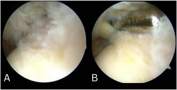
Fig. 1. (A) Confirmation of a partial articular-sided tear of the supraspinatus tendon intraarticularly. (B) Footprint preparation of the supraspinatus tendon using a shaver to enhance bone-to-tendon healing.
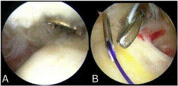
Fig. 2. (A) Progressive dilation is followed by the insertion of the anchor sleeve into the humeral head to facilitate the insertion of both anterior and posterior medial row anchors. (B) Utilization of a spinal needle as a shuttle to secure the suture limbs through the supraspinatus tendon substance, limiting the damage done to the tendon medial to the footprint of the tendon away from the musculotendinous junction.
- Failure of conservative management (Ellman grade 1 and 2).
Note: Considering the aforementioned guidelines, it is crucial for treatment strategies to be individualized, considering factors such as the patient’s activity level, age, and duration of symptoms, in order to achieve optimal outcomes [5].
SURGICAL CONTRAINDICATIONS
- Full-thickness tear of the supraspinatus tendon.
- Intrasubstance or bursal-sided tear of the supraspinatus tendon
- Degenerative tears with poor quality tissue.
TREATMENT OPTIONS
Symptoms associated with partial articular-sided supraspinatus tendon avulsion (PASTA) are often attributed to factors such as pectoral muscle tightness, shoulder capsule tightness, altered movement patterns, and scapular malpositioning rather than solely to the tear itself. In these instances, physiotherapy, nonsteroidal antiinflammatory drugs (NSAIDs), steroid injections, and dynamic stabilization of the scapula and shoulder joint have demonstrated effective pain relief and improved
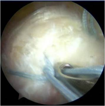
Fig. 3. Tying of the suture limbs using locking sliding knots to promote improved bone-to-tendon contact.
muscle weakness. Consequently, conservative treatment remains the primary approach for tears measuring less than 50% [6].
Among the commonly practiced procedures, three surgical options are frequently employed: 1) Debridement of the footprint alone 2) transtendinous repair (TTR); and 3) completion of tear and subsequent repair (TCR). However, it should be noted that debridement alone does not impede the progression of the tear, so for tears exceeding 50%, transtendinous repair (TTR) and completion of the tear followed by repair (TCR) are commonly recommended modalities [7].
The author recommends utilizing the repair after completion (TCR) technique for elderly patients while advocating for the transtendinous approach in younger patients. This distinction is based on the preserved vascularity typically observed in younger individuals and the need for clearance of degenerative and fatty muscle tissue in older patients with degenerative tears. The tendon takedown procedure during repair provides a conducive environment by creating a foundation of normal tissue that can enhance the healing process [8–10].
OUTCOME OF THE TECHNIQUES
The overall outcome relates to the decrease in the chances of progression of the tear in the supraspinatus tendon and the improvement of healing, which leads to improved functional status for the patient. The meta-analysis done by Yohei et al. showed that the methods of the transtendon technique and repair after completion of the tear showed no difference in the clinical outcome in short- and midterm follow-ups. Also, the retear rates varied from 10% to 90% based on the studies analyzed [5]. However, the meta-analysis done by Sun L et al. that showed
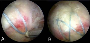
Fig. 4. (A & B) Repair completed with lateral row fixation in a criss-cross configuration of the suture limbs, observed through the subacromial space.
| Transtendinous repair | Tendon takedown procedure |
| Overview of technique | Overview of technique |
| Bursal side of the tendon is kept intact, and repairs are aimed at fixing the footprint of the partially torn tendon to the bony attachment | Repair was undertaken after the completion of the tear from partial to full thickness |
| Pros | Pros |
| Minimal disruption to a healthy supraspinatus tendon | Helps to clear the footprint of supraspinatus tendon attachment and remove degenerate tissues |
| Reduced incidence of retear | Easier footprint preparationfield of view for clearing the Good footprint |
| Cons | Cons |
| Tight folds on the intact surface and uneven tension | More prone for repeat tear |
| Limited area of visualization and needs | Takes down healthy tissues |
more expertise
trans-tendon technique yields better outcomes [9]. The results of long-term outcomes in patients using the transtendinous technique of shoulder arthroscopy have limited data from reviews and meta-analyses to support the overall clinical outcome for the patient. Our patients showed the University of California-Los Angeles (UCLA) shoulder rating scale improvements and returned to sports successfully at the 12-month follow-up, consistent with studies done by Sun L et al. [9].
COMPLICATIONS
The arthroscopic transtendinous approach for repairing PASTA lesions is designed to maintain the integrity of the bursal side of the supraspinatus muscle tendon and to alleviate the symptoms associated [11]. However, it is important to note that during the insertion of anchors for the medial rows, unsuccessful attempts at percutaneous spinal needle insertion can potentially result in damage to the integrity of the bursal surface of the supraspinatus tendon as well as the musculotendinous junction, and failure to do the early postoperative rehabilitation could result in adhesive capsulitis [5].
CONCLUSION AND FUTURE PERSPECTIVES
Expertise is essential for the transtendinous repair technique due to its limited scope for footprint clearance and narrow field of vision. However, mastering this technique is beneficial in preserving the intact fibers of the supraspinatus tendon. In conclusion, proficiency in this technique is crucial for the successful preservation of the supraspinatus tendon’s integrity. Addressing the limited use of anchors for medial footprint attachment and methods to identify over-tensioning in transtendinous repair might improve the overall clinical outcome in young athletes and should be stressed more in future studies.
Ethical approval
No ethical committee approval is required as our presented technique is a modification of an established current technique.
Declaration of competing interests
The authors declare that they have no known competing financial interests or personal relationships that could have appeared to influence the work reported in this paper.
Appendix A. Supplementary data
Supplementary data to this article can be found online at https
://doi.org/10.1016/j.jisako.2024.02.014.
References
- Rossi LA, Ranalletta M. In situ repair of partial-thickness rotator cuff tears: a critical
- analysis review. EFORT Open Reviews 2020;5(3):138–44. https://doi.org/ 10.1302/2058-5241.5.190010.
- Chansky HA, Iannotti JP. The vascularity of the rotator cuff. Clin Sports Med 1991;
- [3] Biberthaler P, Wiedemann E, Nerlich A, Kettler M, Mussack T, Deckelmann S, et al. Microcirculation associated with degenerative rotator cuff lesions. In vivo
assessment with orthogonal polarization spectral imaging during arthroscopy of the
- shoulder. J Bone Joint Surg Am 2003;85(3):475–80. PMID: 12637434.
- Mihata T, McGarry MH, Ishihara Y, Bui CN, Alavekios D, Neo M, et al. Biomechanical
analysis of articular-sided partial-thickness rotator cuff tear and repair. Am J Sports
[10] Med 2015;43(2):439–46. https://doi.org/10.1177/0363546514560156.
- Ono Y, Woodmass JM, Bois AJ, Boorman RS, Thornton GM, Lo IK. Arthroscopic
repair of articular surface partial-thickness rotator cuff tears: transtendon technique
[11] versus repair after completion of the tear-A meta-analysis. Adv Orthop 2016;2016:
7468054. https://doi.org/10.1155/2016/7468054.
- Mihata T, Morikura R, Hasegawa A, Fukunishi K, Kawakami T, Fujisawa Y, et al.
Partial-thickness rotator cuff tear itself did not cause shoulder pain or muscle
weakness in baseball players. Orthop J Sports Med 2019;7(7 suppl 5). https:// doi.org/10.1177/2325967119S00430. 2325967119S00430. Published 2019 Jul 29.
Castricini R, La Camera F, De Gori M, Orlando N, De Benedetto M, Galasso O, et al. Functional outcomes and repair integrity after arthroscopic repair of partial articular supraspinatus tendon avulsion. Arch Orthop Trauma Surg 2019;139:
369–75. https://doi.org/10.1007/s00402-018-3044-4.
Abtahi AM, Granger EK, Tashjian RZ. Factors affecting healing after arthroscopic rotator cuff repair. World J Orthoped 2015;6(2):211–20. https://doi.org/10.5312/ wjo.v6.i2.211.
Sun L, Zhang Q, Ge H, Sun Y, Cheng B. Which is the best repair of articular-sided rotator cuff tears: a meta-analysis. J Orthop Surg Res 2015;10:84. https://doi.org/ 10.1186/s13018-015-0224-6.
Manoharan A, Falgout D, Feldman M. Arthroscopic repair of a PASTA of the shoulder using a bursal split. Arthrosc Tech 2021;10(5):e1403–8. https://doi.org/ 10.1016/j.eats.2021.01.033.
Zhou ZY, Ying H, Wang ZM. Transtendon repair under switching-scope technique for articular partial-thickness rotator cuff tears. Arthroscopy Techniques 2022;11(11):e1973–9. https://doi.org/10.1016/ j.eats.2022.07.013.