

Ayyappan V. Nair a, Maythilisharan Rambhojun a, J. Sreejith Thampy a, Prince Shanavas Khan b,*
- Journal of Orthopaedic Reports – 2024
LINK: https://www.sciencedirect.com/science/article/pii/S2773157X24000067
Cartilage defects of the humeral head are rare and poorly described compared to Hill-Sachs lesions, with limited evidence guiding management. This case report presents the arthroscopic management of an anterosuperior chondral defect in a 43-year-old man with recurrent anterior shoulder instability after a failed Bankart repair. Imaging revealed 15% glenoid bone loss, an off-track Hill-Sachs lesion, and a focal chondral defect of the humeral head.
The patient underwent a single-stage arthroscopic Latarjet, remplissage, and capsulolabral repair, combined with bone marrow aspirate concentrate (BMAC) application to the chondral defect. Bone marrow was aspirated from the iliac crest, processed using the Arthrex Angel system, and applied under dry arthroscopy with CO₂ insufflation. Postoperative rehabilitation included early mobilization, with return to daily activities at 6 weeks and sports at 6 months.
At 1-year follow-up, clinical outcomes showed significant improvement: ASES score increased from 43 to 98 and Rowe score from 30 to 95. CT scans confirmed bony integration, and MRI demonstrated satisfactory filling of both the humeral cartilage and Hill-Sachs defects.
This report highlights the technical feasibility and promising outcomes of arthroscopic BMAC application for humeral head cartilage repair, especially when combined with Latarjet and remplissage in complex instability cases.
1. Introduction Articular cartilage defects of the humeral head are rarer when compared to those of weight bearing joints such as knees and ankles.1 These focal cartilage lesions, which can be a source of pain and disability, can remain elusive on MRI scans and are often only incidentally found during shoulder arthroscopy.2,3 The etiology of these lesions comprises trauma (which can be a single event or repetitive microtrauma), post arthroscopic surgery, intraarticular bupivacaine infusion, osteochondritis dissecans and association with instability and rotator cuff tears.4 The lack of randomized controlled trials is symptomatic of the dearth of high quality evidence that is available in literature to support the clinician in decision making for the management of these cartilage defects.5 Several joint preserving procedures such as debridement, microfracture, OATS (Osteochondral autograft transfer system) and ACI (Autologous chondrocyte implantation), which have stemmed from management of chondral lesions of weight bearing joints, have been adapted and employed in the shoulder.6 However, since the evidence regarding their outcomes is sporadic, inconsistent and of low quality, there is no consensus regarding the superiority of any one of them.4
As reflected in literature, our centre has had good results in using BMACfn1 for the arthroscopic treatment of focal chondral lesions of the knee and ankle,7,8 prompting us to consider this modality in the shoulder. Bone marrow aspirate (BMA) contains mesenchymal stem cells (MSC) that are capable of chondrogenesis and release growth factors that stimulate cartilage repair. Since the concentration of MSCs in unprocessed BMA is merely 0.001%–0.02% of all nucleated cells, density-gradient centrifugation is performed in commercially available systems to increase their concentration by up to 5 times, yielding BMAC.9 The latter is commonly used along with a hyaluronic based scaffold, which has been hypothesized to provide a stable tissue architecture promoting cellular intercations, promoting healing and precluding the use of a periosteal flap cover.7
| * Corresponding author. Department of Orthopaedics, Apollo Adlux Hospital, Angamali, Kochi, Kerala, 683576, India. E-mail address: drpskhan@gmail.com (P.S. Khan). 1 BMAC – Bone Marrow Aspiration Concentrate. https://doi.org/10.1016/j.jorep.2024.100311 Received 21 December 2023; Received in revised form 19 January 2024; Accepted 20 January 2024 Available online 9 February 2024 2773-157X/© 2024 The Author(s). Published by Elsevier B.V. on behalf of Prof. PK Surendran Memorial Education Foundation. This is an open access article under the CC BY-NC-ND license (http://creativecommons.org/licenses/by-nc-nd/4.0/). |
Given the association of focal chondral defects with shoulder instability, the absence of any pertinent literature describing an all arthroscopic technique of chondral repair of humeral head during an arthroscopic Latarjet combined with remplissage procedure spurred our
endeavour to publish this case report.
2. Case report
A 43 years old gentleman presented to our clinic with complaints of recurrence of pain and more than 10 episodes of anterior instability following arthroscopic Bankart repair of the right shoulder 8 years ago at another institution. He could manage only light household activities and pain prevented him from engaging in swimming and playing badminton. He had no complaints in his left, non-dominant shoulder.
The nature of his employment was sedentary (Graphics designer) and he had no comorbidities and no relevant familial history.
3. Clinical findings
On general physical examination, he was moderately built and had a Beighton score of 6/9.
On inspection, both shoulders were level with symmetrical muscle bulk. Healed arthroscopic portals of previous Bankart surgery were noted.
Examination of his right shoulder revealed pain-free full range of movements, at par with the left shoulder in all planes.
No neurological deficits were noted and power was comparable on both sides.
Apprehension test at 90 degrees of shoulder abduction and Jobe’s relocation test were positive whereas sulcus sign was negative.
Drop arm sign, empty can, full can and Hawkins tests were negative.
The preoperative ASES[1] and Rowe scores were 43 and 30 respectively.
4. Diagnostic assessment
4.1. Radiological assessment
The glenoid bone loss was estimated to be 15 per cent with off-track Hill-Sachs lesion from 3D CT[2] scans using the best-fit circle method. MRI[3] scans revealed a Hill-Sachs defect with an anterosuperior cartilage defect of the humeral head (Figs. 1 and 2).
DIAGNOSIS: The patient was diagnosed as having recurrent anterior instability of the right shoulder with critical glenoid bone loss, Hill- Sachs defect with an adjacent chondral defect, status post failed Bankart repair.
4.2. Therapeutic intervention
Considering failed Bankart repair in the past along with critical glenoid bone loss (15%), the patient was counselled for arthroscopic Latarjet procedure along with remplissage, capsulolabral repair and management of the superior cartilage defect with BMAC application at the same sitting.
No ethical committee approval required as our presented technique is a modification of an established current technique.
The arthroscopic Latarjet procedure, during which the coracoid process is osteotomized and transferred to the anterior rim of the glenoid through a subscapularis split, was first described by Lafosse in 2007 (10) and is indicated in anterior instability with critical bone loss of up to 40%, contact athletes and revision cases for failed bankart repair.10 Its advantages over traditional open Latarjet procedure are cosmesis, lesser postoperative fibrosis, ability to address concurrent glenohumeral pathologies and better visualization for accurate coracoid graft positioning.11
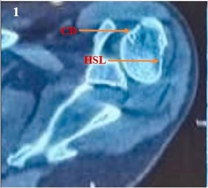
Fig. 1. The image of an axial slice of a plain CT scan of the right shoulder depicting the anterosuperior humeral chondral defect and the posterosuperior Hill-Sachs lesion, labelled as ‘CD’ and ‘HSL’ respectively.
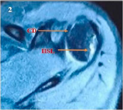
Fig. 2. -Axial T2 weighted MRI image depicting the anterosuperior humeral chondral defect and the posterosuperior Hill-Sachs lesion, labelled as ‘CD’ and ‘HSL’ respectively.
We consider 15% as threshold for critical glenoid bone loss and recommended remplissage procedure due to the presence of an ‘off- track’ Hill-Sachs lesion, in accordance with the recommendations made by Gowd et al. as a result of their systematic review on management of bipolar bone loss in anterior shoulder instability.12
5. Surgical intervention
Informed consent was taken from the patient for the procedure. Arthroscopic Latarjet procedure was performed as described by
Lafosse.13
6. Patient setup and positioning
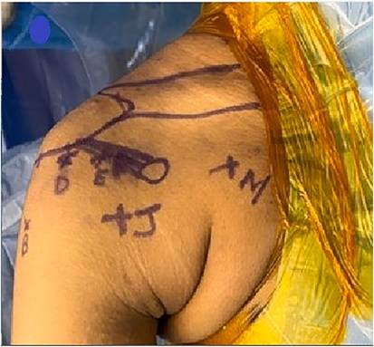
Under general anaesthesia and an interscalene block, the patient is positioned in a beach chair position. 5kg traction is applied to the forearm, keeping the arm elevated at 60◦ with 10 degrees of shoulder abduction and in neutral rotation. The right shoulder was prepped with povidone iodine (7.5%) and draped. The area overlying the right iliac crest till the inguinal region was also prepped in a similar fashion for bone marrow aspiration. 7. Portal placement (Fig. 3)
A portal: 2cm inferior and 2cm medial to the tip of the posterolateral corner of the acromion, corresponding to the soft spot of the standard posterior portal – employed for visualization and introduction of switching stick for splitting the subscapularis.
E portal: Made by outside-in technique through the rotator interval, superior to the lateral half of the subscapularis, used as a working portal for instilling BMAC.
Accessory posterolateral portal: made posterolateral to the posterior portal by outside in technique, used for performing the remplissage.
Trans cuff portal: made at the junction of the musculotendinous junction of the supraspinatus. Used for the visualization of cartilage defect on the anterosuperior humeral head.
D portal: Lateral Portal made anterior to the long head of the biceps. Used for instruments during coracoid preparation and coracoid osteotomy. Also used for visualization during the shoulder’s anterior access, exposure, subscapularis split and fixation of the graft.
H Portal: Anterosuperior portal above coracoid. Used for coracoid drilling and coracoid osteotomy.
J portal: Anteroinferior portal above subscapularis. Used for visualization during the preparation, osteotomy, and transfer of the coracoid graft. Also used for visualization of a superior cartilage defect of the humeral head.
I portal: Ancillary Portal. Used to make the subscapularis split, also used for visualization during the preparation of the coracoid holes.
M portal: Anterior Portal made medial to the conjoint tendon. Used for the pectoralis minor tendon release and double-barrel coracoid positioning cannula.
8. Surgical procedure
Diagnostic arthroscopy performed from the posterior portal revealed earlier placed fiberwire that had been ripped from the labrum. The arthroscope was shifted to the anterolateral trans cuff portal for better visualization of the cartilage defect on the anterosuperior humeral head. Hill-Sachs defect was visualized from the trans cuff portal and remplissage of the infraspinatus tendon was performed using triple- loaded all suture anchors (Stryker, Kalamazoo, MI) working from the accessory posterolateral portal. The suture threads after remplissage were tightened as the final step, after the Latarjet and BMAC procedures. Arthroscopic Latarjet procedure using 3.5mm cannulated titanium metallic screws (DePuy Mitek, Raynham, MA) was performed as described by Lafosse. The focus was shifted to the management of the superior humeral head cartilage defect, which was visualized from the J portal (Fig. 4). A shaving burr was introduced through the anterior E portal to debride the bed of the defect measuring 2 × 2cm and graded as Outerbridge grade 2 cartilage defect (Fig. 5). Bone marrow was aspirated percutaneously from the right iliac crest by inserting the trocar of the bone marrow aspiration kit (Arthrex, Naples, FL) approximately 3 finger breadths proximal to the anterior superior iliac spine. 60 mL of the bone marrow aspirate14 was collected and processed in the Arthrex Angel System (Arthrex, Naples, FL). The prepared cartilage defect area was dried with a sterile swab and to maintain the field dry while producing adequate joint distension, carbon dioxide was insufflation was performed at a pressure of 15 mmHg and at a flow rate of 2L/min, the safety of which had been previously demonstrated in knee arthroscopy.15 The final BMAC concentrate was applied under vision to cover the cartilage defect area working from the anterior E portal (Fig. 6). No further wash was given so the scaffold with the marrow concentrate was converted to a compact concentrate plug.
9. Post operative rehabilitation protocol
Postoperatively, the patient was immobilized in an arm sling. Shoulder shrugs, passive elbow mobilisation and hand grips were started from day one after surgery. Passive shoulder forward flexion and external rotation were started on the 10th day to achieve full passive forward flexion and external rotation at the end of the six weeks. The patient was encouraged to perform computer work after suture removal
Fig. 3. Photo of the draped operative field and the skin marked with the portals described above.
on day 10 after surgery. Active shoulder movements and strengthening
Fig. 4. The anterosuperior chondral defect labelled as ‘CD,’ viewed from the anterolateral trans cuff portal. The long head of biceps is labelled as ‘LHBT.’
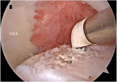
Fig. 5. A radiofrequency ablator through the anterior E portal being used to debride the chondral defect labelled as ‘CD,’ while viewing from the J portal. The glenoid is labelled as ‘GLE.’
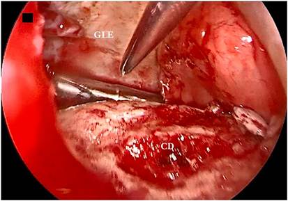
Fig. 6. Similar viewing field as in Fig. 5, post BMAC application over the chondral defect under dry arthroscopy with cardon dioxide insufflation. Chondral defect labelled as ‘CD’ and glenoid as ‘GLE.’
exercises were initiated at the end of six weeks and continued until the third month. The additional procedure did not warrant any deviation from routine rehabilitation for the Latarjet and remplissage procedure. The patient was allowed to return to daily activities in the sixth week and sporting activities were allowed only after six months.
10. Follow-up and outcomes
Clinical outcomes consisted of range of motion assessment while functional outcomes were evaluated using Rowe and ASES (American Shoulder and Elbow Score) scores prospectively up to one year. Radiological evaluation consisted of computed tomography (CT) scans at 3rd and 6th month and magnetic resonance imaging (MRI) at 12th month post operatively to assess bony union and filling of the cartilage defect (Fig. 7).
The ASES score increased from 43 preoperatively to 98 postoperatively while the Rowe score increased from 30 to 95 postoperatively at one year follow up.
CT scans showed good bony integration and remodelling while MRI showed satisfactory filling of the cartilage defect as well as complete filling of the Hill-Sachs defect corresponding to Grade 4 of the Filling Index Score of Remplissage scoring system.
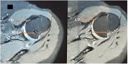
Fig. 7. MRI of the right shoulder one year showing healing of the cartilage defect. Complete filling of the Hill Sachs lesion with good healing of the capsulotenodesis is also noted.
11. Discussion
Focal chondral lesions of the shoulder pose a diagnostic and therapeutic conundrum to the treating clinician. They may be overlooked on MRI as Magnetic Resonance Arthrography has a reported sensitivity of 53–100% and a specificity of 51–87% in detecting humeral chondral lesions, which appear as contour deformities with areas of signal intensity on MRI, typically medial to the Hill-Sachs lesion in case of lesions involving the superior humeral head.2,3 Once their presence is confirmed arthroscopically, they can be treated by several procedures, subdivided into palliative, reparative, restorative and reconstructive (reserved for larger defects).6 Palliative treatment, consisting of debriding the defect, is reserved for small defects in the elderly or in unanticipated lesions, producing a short lived pain relief. Reparative procedures such as microfracturing, subchondral drilling and abrasion chondroplasty induce formation of fibrocartilaginous tissue through bone marrow stimulation.6 Microfracture is the most commonly used technique and can be combined with other reparative and restorative procedures.5 Millett et al. reported failure of 3 out of 12 cases of full thickness chondral lesions affecting the humeral head after microfracture in their case series.16 Unlike reparative procedures, restorative procedures like OATS, ACI and BMAC aim at repairing the defect with hyaline-like tissue. Comparative studies in shoulder, albeit sparse, have demonstrated superiority of BMAC over microfracture and similar outcomes of the former with matrix-induced ACI.17,18 In the knee, however, BMAC has a proven track record in the treatment of chondral lesions, reportedly improving Visual Analogue Score from 5.4 to 0.5 and International Knee Documentation Committee Subjective Knee Form score from 39.2 to 82.2. Interestingly, follow up MRI showed excellent filling of defect in 81% of patients under 45 years of age and 70% of those older than 45 years.19 Similarly, when used in talar chondral lesions, MRI revealed 77.3% defects were completely filled and significant improvement in Foot and Ankle Outcome Score was reported.20 Given the supportive evidence for the use of BMAC in treating chondral defects of these weight bearing joints, we anticipated at least equally good results in the shoulder in a single stage and at a lower cost compared to ACI. The excellent functional and radiological outcomes in this patient justify the use of BMAC in treating chondral defects of the humeral head. 12. Limitation of study
Complications like arthritis could not be assessed and we could not opine on whether the excellent functional outcomes were sustained in the long term due to the one year follow up period.
Financial support and sponsorship
No financial support or sponsorship was solicited nor received for this case.
Ethical statement
This Case report had no objection from the institutional research Committee and was submitted after the IRB approval.
Funding
This study has not received any remuneration or support for the financial disclosure.
Patient consent
This Case report is being published after obtaining informed consent from the patient.
CRediT authorship contribution statement
Ayyappan V. Nair: All Authors have contributed to the work in this journal. Maythilisharan Rambhojun: was the principal investigator and the first author and was involved in data collection, statistical analysis, observation. J. Sreejith Thampy: involved in manuscript editing services, proof reading along with. Prince Shanavas Khan: was monitoring the progress in the research discussions and was the mentor for writing.
Declaration of competing interest
The authors report no conflict of interest.
Appendix A. Supplementary data
Supplementary data to this article can be found online at https://doi.
org/10.1016/j.jorep.2024.100311.
References
- Løken S, Granan LP, Sivertsen EA, Årøen A. Epidemiology of cartilage injuries. In: Sports Injuries. Berlin, Heidelberg: Springer Berlin Heidelberg; 2013:1–12.
- Carroll KW, Helms CA, Speer KP. Focal articular cartilage lesions of the superior humeral head. Am J Roentgenol. 2001 Feb;176(2), 393–7.
- Guntern DV, Pfirrmann CWA, Schmid MR, et al. Articular cartilage lesions of the glenohumeral joint: diagnostic effectiveness of MR Arthrography and prevalence in patients with subacromial impingement syndrome. Radiology. 2003 Jan;226(1),
- Dagher D, Selznick A, Prada C, Al Shehab Y, Leroux T, Khan M. Surgical treatment options for articular cartilage defects of the glenohumeral joint: a systematic review. Shoulder Elbow. 2023 Dec 14;15(6):580–592.
- Fiegen A, Leland DP, Bernard CD, et al. Articular cartilage defects of the glenohumeral joint: a systematic review of treatment options and outcomes. Cartilage. 2021 Dec 23;13(1_suppl):401S–413S.
- Gomoll AH, McCulloch P, Kang RW, Cole BJ. Treatment of chondral defects in the shoulder. Operat Tech Orthop. 2006 Oct;16(4):232–243.
- Chahla J, Dean CS, Moatshe G, Pascual-Garrido C, Serra Cruz R, LaPrade RF. Concentrated bone marrow aspirate for the treatment of chondral injuries and osteoarthritis of the knee. Orthop J Sports Med. 2016 Jan 1;4(1), 232596711562548.
- Lan T, McCarthy HS, Hulme CH, Wright KT, Makwana N. The management of talar osteochondral lesions – current concepts. J Arthrosc Joint Surg. 2021 Jul;8(3):
- Dragoo JL, Guzman RA. Evaluation of the consistency and composition of commercially available bone marrow aspirate concentrate systems. Orthop J Sports Med. 2020 Jan 1;8(1), 232596711989363.
- Moya D, Aydin N, Yamamoto N, et al. Current concepts in anterior glenohumeral instability: diagnosis and treatment. SICOT J. 2021 Sep 14;7:48.
- Haskel JD, Colasanti CA, Hurley ET, Matache BA, Jazrawi LM, Meislin RJ. Arthroscopic Latarjet procedure. JBJS Rev. 2021 Mar;9(3).
- Gowd AK, Liu JN, Cabarcas BC, et al. Management of recurrent anterior shoulder instability with bipolar bone loss: a systematic review to assess critical bone loss amounts. Am J Sports Med. 2019 Aug 27;47(10):2484–2493.
- Lafosse L, Lejeune E, Bouchard A, Kakuda C, Gobezie R, Kochhar T. The arthroscopic Latarjet procedure for the treatment of anterior shoulder instability. Arthrosc J Arthrosc Relat Surg. 2007 Nov;23(11):1242.e1–1242.e5.
- Davila Castrodad IM, Mease SJ, Werheim E, McInerney VK, Scillia AJ. Arthroscopic´ chondral defect repair with extracellular matrix scaffold and bone marrow aspirate concentrate. Arthrosc Tech. 2020 Sep;9(9):e1241–e1247.
- Penton JL, Flick TR, Savoie FH, Heard WM, Sherman WF. Midterm safety of carbon dioxide insufflation of the knee during arthroscopic cartilage–based procedures. Orthop J Sports Med. 2021 Oct 1;9(10), 232596712110354.
- Millett PJ, Huffard BH, Horan MP, Hawkins RJ, Steadman JR. Outcomes of full- thickness articular cartilage injuries of the shoulder treated with microfracture. Arthroscopy. 2009 Aug;25(8):856–863.
- Cotter EJ, Wang KC, Yanke AB, Chubinskaya S. Bone marrow aspirate concentrate for cartilage defects of the knee: from bench to bedside evidence. Cartilage. 2018 Apr 10;9(2):161–170.
- Cavinatto L, Hinckel BB, Tomlinson RE, Gupta S, Farr J, Bartolozzi AR. The role of bone marrow aspirate concentrate for the treatment of focal chondral lesions of the knee: a systematic review and critical analysis of animal and clinical studies. Arthroscopy. 2019 Jun;35(6):1860–1877.
- Gobbi A, Scotti C, Karnatzikos G, Mudhigere A, Castro M, Peretti GM. One-step surgery with multipotent stem cells and Hyaluronan-based scaffold for the treatment of full-thickness chondral defects of the knee in patients older than 45 years. Knee Surg Sports Traumatol Arthrosc. 2017 Aug 14;25(8):2494–2501.
- Hannon CP, Ross KA, Murawski CD, et al. Arthroscopic bone marrow stimulation and concentrated bone marrow aspirate for osteochondral lesions of the talus: a case-control study of functional and magnetic resonance observation of cartilage repair tissue outcomes. Arthrosc J Arthrosc Relat Surg. 2016 Feb;32(2):339–347.
[1] ASES – American Shoulder and Elbow Society.
[2] CT – Computed Tomography.
[3] MRI – Magnetic Resonance Imaging.