

Jimmy Joseph Meleppuram, M.S.Ortho., Ayyappan V. Nair, D.Ortho., D.N.B.Ortho., Hamdi Nizar Ahamed, M.S.Ortho., Nizaj N, D.N.B.Ortho., M.R.C.S.(Glasgow),
Aebel Raju, M.B.B.S., M.R.C.S.(England), Ajayakumar Thankappan, M.S.Ortho., and Prince Shanavas Khan, D.Ortho., M.S.Ortho.
| P |
osterior ankle impingement syndrome causes sharp posterior ankle pain during plantar flexion, often seen in athletes.1-3 Although early cases may respond to conservative treatment, 40% of patients require surgery. Posterior ankle arthroscopy is a minimally invasive option, particularly effective for os trigonum syndrome, in which repetitive trauma or injury to the os trigonum leads to persistent hindfoot pain4-6 (Figs 1 and 2).
Posterior ankle arthroscopy has replaced open surgery for symptomatic os trigonum because of its minimally invasive nature, offering precise removal with less tissue disruption, reduced pain, fewer complications, and faster recovery. It also allows simultaneous treatment of other pathologies, enhancing outcomes and enabling a quicker return to sports, making it the preferred option for athletes.7 The
advantages and disadvantages of the procedure are presented in Table 1.
Surgical Technique
Setup
Under spinal anesthesia, the patient is positioned prone, with care taken to cushion pressure-prone areas (Video 1). The ankle is left dangling off the table for unrestricted movement. A thigh tourniquet is applied to allow a clear surgical field. Pre-incision marking is performed, with portals planned using anatomic landmarks. The posterolateral (PL) portal is created just above the intermalleolar line, 1 mm lateral to the Achilles tendon, and the posteromedial (PM) portal is placed at the same level medially.8,9 A longitudinal line along the lateral border of the third metatarsal is marked on the plantar surface to guide instrument trajectory and minimize medial neurovascular injury risk (Fig 3).
Posterior Ankle Arthroscopy
After inflation of the tourniquet, 10 mL of lignocaineepinephrine (4 mL) with saline solution (6 mL) is injected into the PL and PM portals via a 23-gauge needle before incision. The subcutaneous tissue and deeper layers are dissected bluntly using a straight hemostat clamp, oriented in line with the lateral border of the third metatarsal. A 4.0-mm 30 arthroscope (Smith & Nephew) is then introduced into the PL portal. The trocar connects to a closed pump at 30 to 40 mm Hg.
Arthroscopy Techniques, Vol 14, No 6 (June), 2025: 103521 e1
e2 J. J. MELEPPURAM ET AL.
Fig 1. Preoperative radiographs of right ankle in male rig worker with chronic recurring posterior ankle pain and elicited tenderness after sustaining football injury, aggravated by work-related forced plantar flexion and dorsiflexion. Anteroposterior and lateral views depict the os trigonum with smooth borders, mainly seen on the lateral view. (R, right.)
The vertical PM portal incision is made parallel to the PL portal at the same level in a similar manner under needle guidance, viewing from the PL portal. Deep dissection is carried out with a mosquito clamp, which is directed toward the arthroscope shaft at a 90 angle. The clamp is then maneuvered along the shaft and opened in front of the arthroscope tip to create space and enhance visualization. Dense fibrotic and fatty tissues are commonly encountered posterior to the talus initially, potentially hindering visualization. A 2.9-mm full-radius shaver is inserted into the PM portal to perform initial soft-tissue debridement and exposure of the intermalleolar ligament in the superolateral quadrant. The posterior ankle joint lies superior to the intermalleolar ligament, whereas the subtalar joint is located inferior to it. The aforementioned portals are interchanged for working as well as visualization as required by the surgeon’s preference (Fig 4).9,10 The intermalleolar ligament might be hypertrophied and is found to impinge on the surrounding tissues, which are eventually debrided with a shaver or punch. The surgeon should assess and debulk the transverse and
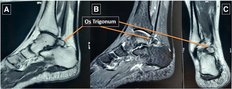
Fig 2. Magnetic resonance imaging scans of right ankle: T1-weighted (A) and short tau inversion recovery (B) sagittal views confirming os trigonum with adjacent bone marrow edema, without avascular necrosis, and T1-weighted coronal view (C) depicting os trigonum in inferomedial quadrant. (PLS, posterior lateral superior; RPI, right posterior inferior; SRA, superior right anterior; SPR, superior posterior right.)
ARTHROSCOPIC EXCISION OF OS TRIGONUM e3
Table 1. Advantages and Disadvantages of Posterior Ankle Arthroscopy
Advantages
Decreased wound complications, insignificant blood loss, better cosmesis, and potential for quicker recovery
Less invasive and able to be performed in wide-awake theater scenarios
Direct visualization for diagnostic purposes far superior to MRI
Does not require distraction device and can be performed with usual 4-mm 30 scope Reduced cost
Disadvantages
Cannot be performed in office/outpatient setting
Difficult to convert to open procedure if required Learning curve
MRI, magnetic resonance imaging.
posterior inferior tibiofibular ligaments if impingement is present.
The flexor hallucis longus (FHL) tendon is an essential landmark and must be identified, debrided from its sheath, and kept in view medially throughout the procedure to prevent iatrogenic damage to the neurovascular bundle in the superomedial quadrant. The FHL tendon must be evaluated for tenosynovitis, a low-line muscle belly, and tethering. Passive plantar flexion and dorsiflexion of the great toe confirm and demonstrate impingement of the FHL tendon against the adjacent hypertrophic intermalleolar ligament, making the procedure both streamlined and effective (Fig 5).
The arthroscope is directed inferiorly to the inferomedial quadrant to visualize the os trigonum, and the os trigonum is demarcated in its entirety, if possible, by removing all the soft tissues attached to it. The synchondrosis, or cartilage connection, between the PL talar process and the os trigonum is carefully separated through levering maneuvers; otherwise, it is taken out in pieces using a grasper or burred off in toto (Fig 6).
Finally, the surgeon evaluates the inferolateral quadrant for any pathologies and, if required, specif-
ically addresses these pathologies as appropriate. The surgeon evaluates the talus, resects the capsule, and smoothens sharp edges in joints. Osteochondral lesions, synovitis, osteophytes, hypertrophic capsules, and impingement should be identified and treated. Osteochondral defects and subchondral cysts can be debrided and addressed with microfracture or drilling techniques to promote fibrocartilage healing.
Postoperatively, the os trigonum excision is confirmed via fluoroscopy, the wounds are sutured and dressed, and the limb is immobilized with a wellmolded below-knee slab with pillow elevation. The limb is immobilized in the below-knee slab for 2 weeks until suture removal, with noneweight-bearing gait training initiated on postoperative day 1. Full weight
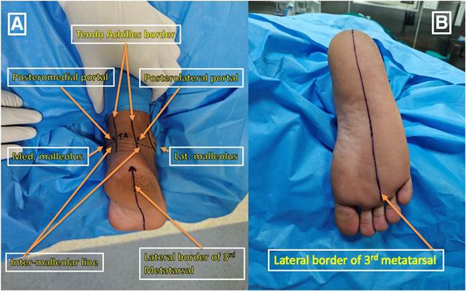
Fig 3. (A) Surface marking of posterolateral and posteromedial portals with intermalleolar line and tendo-Achilles medial and lateral borders in right foot. (B) Marking of lateral border of third metatarsal in right foot on end-on-view. The patient is in the prone position, and the right foot and ankle are dangling off the operating table after elevation with no joint distractor. (Lat, lateral; Med, medial.)
e4 J. J. MELEPPURAM ET AL.
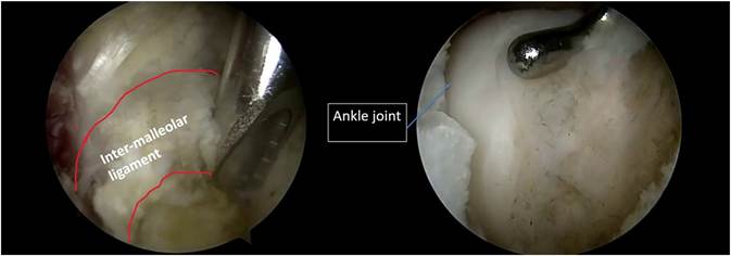
Fig 4. Visualization using 4-mm 30 arthroscope from posteromedial portal and posterolateral portal interchanged per surgeon’s choice. Initial debridement is performed using a 2.9-mm shaver for the intermalleolar ligament, followed by identification of the right posterior ankle joint space distracted by manual traction on the patient’s ankle in the prone position.
bearing is started after suture removal at 2 weeks, and the patient is advised to resume normal activities after 1 month.
Discussion
Posterior ankle impingement syndrome is a condition marked by pain at the back of the ankle, particularly during forced plantar flexion. This pain can be either acute, occurring after trauma, or chronic, occurring due to repetitive stress. The most common cause is pathology involving the os trigonum or talar process. Still, the syndrome can also arise from conditions such as FHL tenosynovitis, ankle osteochondritis, subtalar joint
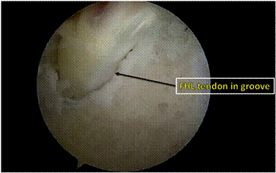
Fig 5. With the patient in the prone position, viewing from the posterolateral portal, visualization of the right flexor hallucis longus (FHL) tendon with its musculotendinous juncture in the groove is confirmed by passively plantar flexing the right great toe during the procedure. It should be noted that no posterior ankle arthroscopy procedure should be performed medially toward the FHL tendon space to prevent damage to neurovascular structures.
disease, or fracture. Patients often experience chronic or recurring posterior ankle pain, especially during activities that involve forced plantar flexion or push-off motions, such as dancing (en pointe toe position), kicking, or downhill running. The diagnosis primarily relies on clinical history and physical examination findings, with radiography, computed tomography, and magnetic resonance imaging used to identify related bone and soft-tissue abnormalities. Surgery is required when conservative management fails to improve
symptoms.3,5,7
The potential complications of the described procedure include plantar numbness, sural nerve dysesthesia, tightening of the Achilles tendon, surgical site infection, and complex regional pain syndrome. Pearls and pitfalls of posterior ankle arthroscopy are presented in
Table 2.11,12
In conclusion, posterior ankle impingement syndrome, often due to os trigonum, impacts quality of life. Our experience with posterior ankle arthroscopy for os trigonum excision highlights its effectiveness, offering reduced pain, minimal scarring, and faster recovery compared with open surgery. The procedure allows precise os trigonum removal while preserving tissues and addressing other intra-articular issues.
Declaration of Generative AI and AIAssisted Technologies in the Writing Process
During the preparation of this work, the authors used free text-to-speech online software (Microsoft clipchamp) to provide background narration for Video 1. After using this tool/service, the author reviewed and edited the content as needed and takes full responsibility for the content of the publication.
| ARTHROSCOPIC EXCISION OF OS TRIGONUM e5 |
FHL, flexor hallucis longus.
Disclosures
All authors (J.J.M., A.V.N., H.N.A., N.N., A.R., A.T., P.S.K.) declare that they have no known competing financialinterestsorpersonalrelationshipsthatcouldhave appeared to influence the work reported in this paper.
References
- Lavery KP, McHale KJ, Rossy WH, Theodore G. Ankle impingement. J Orthop Surg Res 2016;11:46.
- Kembhavi R, Chitten JJ, Khatri RY. Arthroscopic decompression of chronic posterior ankle impingement syndrome provides a good functional outcome. J Orthop Case Rep 2024;14:214-219.
- Ishibashi MA, Doyle MD, Krcal CE Jr. Posterior ankle impingement syndrome. Clin Podiatr Med Surg 2023;40: 209-222.
- McAlister JE, Urooj U. Os trigonum syndrome. Clin Podiatr Med Surg 2021;38:279-290.
- van Dijk CN, van Bergen CJ. Os trigonum syndrome: Diagnosis and management. Oper Tech Orthop 2010;20:46-51.
- Robinson P, White LM. Soft-tissue and osseous impingement syndromes of the ankle: Role of imaging in diagnosis and management. Radiographics 2002;22: 1457-1469.
- MolloyA,Tzortziou-BrownV.Posteriorankleimpingement syndrome in athletes. Clin Sports Med 2015;34:449-460.
- Nickisch F, Barg A, Saltzman CL, et al. Posterior ankle and hindfoot arthroscopy. JBJS Essent Surg Tech 2012;2:e15.
- Gasparetto F, Collo G, Pisanu G, et al. Posterior ankle and subtalar arthroscopy: Indications, technique, and results. Curr Rev Musculoskelet Med 2012;5:164-170.
- Butler JJ, Roof M, Montgomery SR Jr, et al. Basics of ankle arthroscopy part 4: Surface anatomy, portal placement, and diagnostic evaluation for posterior ankle arthroscopy. Arthrosc Tech 2025;14:103323.
- Ogut T, Ayhan E, Irgit K, Sarikaya AI. Posterior ankle impingement: Results of arthroscopic treatment. Arthroscopy 2012;28:711-715.
- Dias CGP, Mansur NSB, Asaumi ID, Lemos AVKC,
Azevedo RM, Nery CA. Severe complications of posterior ankle arthroscopy. Sci J Foot Ankle 2019;13:961.