

Jimmy Joseph Meleppuram, M.S.Ortho., Ayyappan V. Nair, D.Ortho., D.N.B.Ortho.,
Hamdi Nizar Ahamed, M.S.Ortho., Nizaj Nasimudeen, D.N.B.Ortho., M.R.C.S.,
Ananthakrishnan Radhakrishnan, M.B.B.S., Ajayakumar Thankappan, M.S.Ortho., and Prince Shanavas Khan, D.Ortho., M.S.Ortho.
- Arthroscopy Techniques – Jun 2025
LINK: https://pubmed.ncbi.nlm.nih.gov/40656690/
Poliomyelitis, though rare today, continues to cause long-term musculoskeletal deformities, particularly equinocavovarus deformity of the ankle due to muscle imbalance and weakness. If untreated, these deformities lead to abnormal gait, increased energy expenditure, and end-stage ankle arthritis. Arthroscopic ankle arthrodesis has emerged as a minimally invasive option for such cases, offering advantages over open surgery, including reduced soft-tissue trauma, faster recovery, and fewer complications.
The article describes a case of a 53-year-old woman with post-polio residual paralysis and painful ankle deformity who underwent arthroscopic ankle fusion. Using anteromedial and anterolateral portals, arthroscopy revealed cartilage loss at the tibial plafond. Cartilage was removed until subchondral bleeding was achieved, and the ankle was aligned in neutral dorsiflexion, slight eversion, and external rotation. Three converging compression screws were placed under fluoroscopic guidance to achieve stable fixation. Postoperatively, the patient progressed from non–weight-bearing immobilization to gradual physiotherapy and full activities by 12 weeks, with successful pain relief and improved alignment.
Arthroscopic ankle fusion provides high fusion rates with less morbidity compared with open fusion but requires advanced skills, specialized equipment, and is limited in severe deformities or extensive bone loss. Overall, it represents a promising option for post-polio patients with end-stage ankle arthritis.
| A |
lthough poliomyelitis is now rare in developed countries, its long-term effects continue to affect many individuals, especially in developing regions.1 Deformities around the ankle in post-polio patients arise from muscle imbalances, poor posture, and the strain of prolonged walking on a mal-aligned foot. If untreated, these deformities can lead to abnormal antalgic gait, increased energy expenditure, and
eventually, joint arthritis.2-4
Equinocavovarus deformity, common in patients with post-polio residual paralysis, is characterized by a combination of ankle equinus and varus deformity, midfoot cavus, and forefoot clawing or hammertoe deformity. The deformity primarily stems from muscle weakness and imbalance between the extrinsic and intrinsic muscles. An overactive peroneus longus compensates for a weak tibialis anterior, leading to hypereplantar flexion of the first metatarsal. This forces the long toe extensors to overcompensate, worsening
midfoot cavus. End-stage ankle arthritis is a common outcome of untreated or poorly managed deformities in post-polio patients.2,3 Arthroscopic assisted ankle arthrodesis as performed for medial tibial plafond arthritic changes in bones that are both small in variation and soft in a post-polio patient offers hope through precise correction of deformities and minimal invasiveness, resulting in faster recovery and reduced complications compared with open surgery3,4 (Figs 1 and 2).
Surgical Technique
Setup
Under spinal anesthesia, the patient is positioned supine with a bump under the ipsilateral hip to place the foot in a neutral, pressure-free position (Video 1). A pneumatic tourniquet is applied at the appropriate pressure. The ankle to be operated on is carefully positioned to dangle off the edge of the table, enabling free movement throughout the procedure. After sterile preparation anddrapingofthe operative extremity, with the use of a sterile marking pen, the relevant anatomic landmarks are marked, including the lateral malleolus, medial malleolus, anteromedial portal, anterolateral portal,5 and superficial peroneal nerve depicted by the fourth-toe flexion sign or Stephen sign6 (Fig 3).
Arthroscopic Ankle Fusion
After tourniquet inflation, a 23-gauge needle injects 10 mL of lignocaine-epinephrine (4 mL) with saline solution (6 mL) into the anteromedial and anterolateral
Arthroscopy Techniques, Vol 14, No 6 (June), 2025: 103547 e1
Fig 1. Supine anteroposterior (AP) and lateral (LAT) radiographic views of involved left ankle showing medial tibial plafond arthritic (minimal) changes along with smallsized bone.
portals before incision. The anteromedial portal is created at the joint line, medial to the tibialis anterior tendon in the soft spot palpated after dorsiflexion and plantar flexion of the foot, and a 4.0-mm, 30 arthroscope (Smith & Nephew, Andover, MA) is introduced, followed by anterolateral portal placement just lateral to the peroneus tertius tendon, with care taken to protect the superficial peroneal nerve. Diagnostic arthroscopy reveals minimal narrowing of the medial tibial plafond joint space, as well as denuded cartilage at the notch of Harty. The bones were found to be relatively small and soft5-7 (Fig 4).
Further evaluation carefully assesses for the presence of any osteophytes, loose bodies, or osteochondral lesions of the talus and tibial plafond. A 3-mm shaver (Arthrex, Naples, FL) is used to debride interposing soft tissue to aid clarified vision. All the remaining articular cartilage is denuded with the shaver (ring curette [Arthrex] or osteotomes may be used), and lavage is performed in the joint and outlet pathway until subchondral bleeding spots are seen both in the talar dome and under the tibial articulating surface (Fig 5). Another key indicator of reaching the right depth is the appearance of fat bubbles emerging fromtheboneonceirrigationstops.Reductionoftheankle isheldandmaintainedmanuallyinneutraldorsiflexion,5 of eversion, and 10 of external rotation using K-wires. Then, 3 guidewires for 6.5-mm compression screws (Osteoplus; Sorath Ortho, Ahmedabad, Gujarat, India), initially pointing from the centro-medial distal tibia to the anterolateral talar head and neck, followed by pointing fromtheposterolateraldistaltibiatotheanteromedialtalar head and neck across the tibiotalar joint and, finally, pointing from the posteromedial distal tibia to the anterolateral talar neck, are placedin a converging mannervia anteroposterior- and lateral-view fluoroscopic images (Figs 6-8). After making the necessary skin incisions, the surgeon performs measurements as well as drilling, and proceeds with screw placement, making sure to verify good purchase and compression of the tibiotalar joint surface with no screw extrusion into the subtalar joint surface. After satisfactory screw placement is achieved, the guidewires are removed and final images are obtained (Fig 9). Closure of the skin is achieved with No. 3-0 nylon. The wound sites are dressed, and the patient is
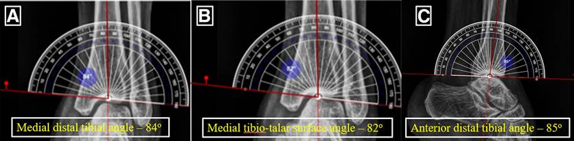
Fig 2. (A) An anteroposterior weight-bearing view of the left ankle shows the medial distal tibial angle, formed on the medial side by the mechanical axis of the tibia and a tangent drawn along the tibial plafond, measuring 84, signifying minimal varus. (B) An anteroposterior weight-bearing view of the left ankle shows the medial tibiotalar surface angle, formed on the medial side by the mechanical axis of the tibia and a tangent drawn along the talar dome, measuring 82, signifying minimal varus. (C) A lateral weight-bearing view of the left ankle shows the anterior distal tibial angle, formed from the anatomic axis of the tibia and the line connecting distal points on the anterior and posterior tibial articular surface, measuring 85, signifying minimal equinus mispositioning of talus.
ARTHROSCOPIC FUSION IN POST-POLIO FOOT e3
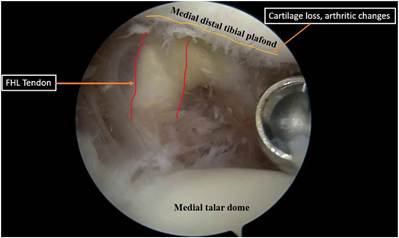
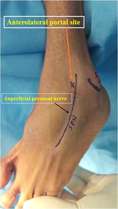
Fig 3. Superficial peroneal nerve (SPN) depicted by fourthtoe flexion sign (Stephen sign) in left ankle in supine position as foot dangles free of table.
immobilized with a below-knee slab in neutral dorsiflexion and slight eversion and external rotation with pil-
low elevation.5,6,8
Postoperative Protocol
The postoperative protocol is as follows: At 0 to 2 weeks, the patient is noneweight bearing, receives antibiotics, and uses analgesics as well as ice packs for pain management. At 2 to 6 weeks, the patient undergoes wound inspection, suture removal, and cast conversion with continued noneweight bearing. At 6 to 8 weeks, progressive weight bearing is initiated and the patient begins physical therapy with range-ofmotion exercises. The patient progresses to more weight-bearing activities and functional exercises at 8
Fig 4. Arthroscopic inlet view of left ankle in supine position via anterior portal visualizing medial notch of Harty with minimal arthritic changes due to cartilage loss and anterior aspect of flexor hallucis longus (FHL) tendon in background demarcated with medial distal tibial plafond superiorly as well as medial talar dome inferiorly.
to 12 weeks. Finally, the patient advances to more vigorous activities, depending on his or her recovery, at 12 weeks onward.6
Discussion
Poliomyelitiscausesmuscleweakness duetodamageto theanteriorhorncellsinthespinalcord,leadingtomuscle imbalances and joint deformities. End-stage ankle arthritis, due to post-polio residual paralysis, is a debilitating condition that severely impairs daily function and quality of life.1,3 Arthroscopic tibiotalar fusion is the preferred treatment for end-stage ankle arthritis unresponsive to conservative management. It offers several advantages, including reduced soft-tissue damage, faster recovery, and lower complication rates.9 In thepresented case,a53-year-oldfemalepatientwithpost-polioresidual paralysis presented with severe pain and deformity of the left ankle. After a thorough preoperative evaluation, she
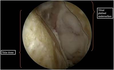
Fig 5. Arthroscopic inlet view of left ankle in supine position with anterior-portal visualization showing completely denuded articular cartilage with subchondral bleeding spots seen both in talar dome and under surface of tibial plafond.
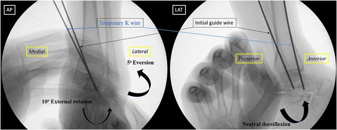
Fig 6. Fluoroscopic anteroposterior (AP) and lateral (LAT) images of left ankle under manual reduction held and maintained in neutral dorsiflexion, 5 of eversion, and 10 of external rotation using K-wire followed by initial guidewire for 6.5-mm compression screw insertion from centro-medial distal tibia to anterolateral talar neck direction.
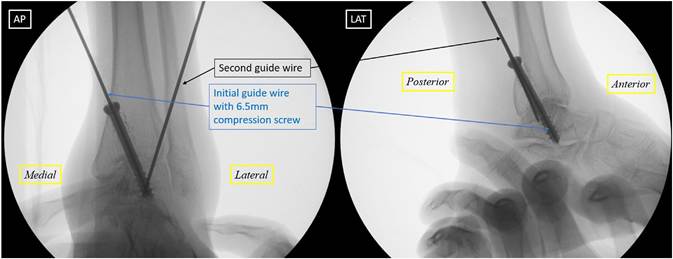
Fig 7. Fluoroscopic anteroposterior (AP) and lateral (LAT) views of left ankle held in manual traction and reduction. The second guidewire for the compression screw is inserted from the posterolateral distal tibia to the anteromedial talar head direction.
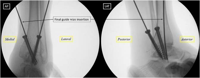
Fig 8. Fluoroscopic anteroposterior (AP) and lateral (LAT) views of left ankle held in manual traction and reduction. The final guidewire for the compression screw is inserted from the posteromedial distal tibia to the anterolateral talar neck in converging fashion with the initial and second 6.5-mm compression screws in situ.
underwentarthroscopicassistedanklearthrodesis,which Despite its benefits, arthroscopic ankle arthrodesis resulted in successful fusion, improved alignment, and carries risks, including incomplete denudation, poor significant pain reduction. bone quality, and implant failure. Complications may
ARTHROSCOPIC FUSION IN POST-POLIO FOOT e5
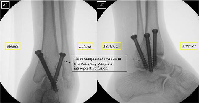
Fig 9. Final fluoroscopic images of fusion in anteroposterior (AP) and lateral (LAT) views of left ankle illustrating three 6.5-mm compression screws, initially pointing from centro-medial distal tibia to anterolateral talar head and neck, followed by pointing from posterolateral distal tibia to anteromedial talar head and neck across tibiotalar joint and, finally, pointing from posteromedial distal tibia to anterolateral talar neck, placed in converging manner.
Table 1. Advantages and Disadvantages of Arthroscopic Ankle Arthrodesis
| Aspect | Advantages | Disadvantages |
| Surgical approach | Minimally invasive with smaller incisions, reduced scarring, and better cosmetic results | Requires advanced surgical skills and specialized equipment |
| Recovery time | Faster recovery and shorter hospital stay | Limited visualization in complex deformities compared with open techniques |
| Postoperative pain | Less postoperative pain owing to minimal soft-tissue dissection | May not be suitable for patients with severe deformities or large bone defects |
| Infection risk | Lower risk of wound infection and soft-tissue complications | Potential risk of joint infection through small portals, though rare |
| Alignment accuracy | High accuracy in achieving joint alignment with fluoroscopic guidance | Difficulty in achieving alignment in cases of severe deformity or malalignment |
| Functional outcomes | Comparable or better outcomes in terms of pain relief and fusion rates vs open fusion | Limited access in cases with significant bone loss or hardware removal needs |
| Operative time | Typically shorter than open surgery | Can be prolonged in complex cases or if technical difficulties arise |
| Cost | Lower overall cost owing to reduced hospitalization and complications | Initial investment in arthroscopic equipment can be high |
| Table 2. Comparison of Open Versus Arthroscopic Ankle Arthrodesis | ||
| Aspect | Open Ankle Arthrodesis | Arthroscopic Ankle Arthrodesis |
| Incisions | Large incisions, leading to more scarring | Smaller incisions with better cosmetic outcomes |
| Soft-tissue dissection | Extensive soft-tissue dissection, increasing tissue damage | Minimal soft-tissue disruption |
| Postoperative pain | Higher postoperative pain due to greater tissue trauma | Reduced postoperative pain |
| Infection risk | Higher risk of infection and wound complications | Lower risk of infection |
| Visualization | Better visualization of joint and surrounding structures in complex deformities | Limited visualization, especially in severe deformities |
| Bone preparation | More accessible for large deformities or extensive bone work | Challenging in cases of severe deformities or significant bone loss |
| Operative time | Can be longer in some cases because of more extensive procedures | Generally shorter but can be prolonged in complex cases |
| Alignment accuracy | Effective in correcting severe deformities and malalignment | Effective for mild to moderate deformities; challenging in severe cases |
| Recovery time | Longer recovery period with increased hospital stay | Faster recovery and shorter hospital stay |
| Fusion rate | High fusion rate but comparable to arthroscopic fusion | High fusion rate with less morbidity |
arise from improper portal placement or neurovascular injury. Proper training in ankle arthroscopy and comprehensive postoperative care are essential to optimize outcomes and reduce complications.10 Generally, contraindications for arthroscopic ankle arthrodesis include significant malrotation and bone loss, failed previous fusion, active infection, and significant concomitant subtalar and/or hindfoot joint arthritis. Advantages and disadvantages of arthroscopic ankle arthrodesis are presented in Table 1, and pros and cons of arthroscopic ankle arthrodesis compared with open ankle arthrodesis are depicted in Table 2.11-13
Overall, arthroscopic ankle arthrodesis is a promising option for managing end-stage ankle arthritis in patients with post-polio residual paralysis. Its minimally invasive nature allows for precise deformity correction, faster recovery, and improved quality of life. However, the success of this procedure relies on meticulous surgical technique, appropriate training, and diligent postoperative management to minimize complications.
Declaration of Generative AI and AI-Assisted Technologies in the Writing Process
During the preparation of this work, the authors used free text-to-speech online software, Microsoft Clipchamp, to provide background narration for Video 1. After using this tool/service, the author reviewed and edited the content as needed and take full responsibility for the content of the publication.
Disclosures
All authors (J.J.M., A.V.N., H.N.A., N.N., A.R., A.T., P.S.K.) declare that they have no known competing financialinterestsorpersonalrelationshipsthatcouldhave appeared to influence the work reported in this paper.
References
- Baliga S, McMillan T, Sutherland A, Sharan D. The prevalence and severity of joint problems and disability in patients with poliomyelitis in urban India. Open Orthop J 2015;9:204-209.
- Joseph B, Watts H. Polio revisited: Reviving knowledge and skills to meet the challenge of resurgence. J Child Orthop 2015;9:325-338.
- Shah R, Sharma S, Shah SR. Management of rigid postpoliomyelitis equinocavovarus deformities: A brief review. J Foot Ankle Surg (Asia Pac) 2022;9:135-141.
- Dhillon MS, Sandhu HS. Surgical options in the management of residual foot problems in poliomyelitis. Foot Ankle Clin 2000;5:327-347.
- Mangwani J, Afifi H, Faroug R. Arthroscopic ankle arthrodesis: Surgical technique. J Arthrosc Surg Sports Med 2021;2:135-140.
- Maharjan L, Washburn F, Tran B, Pyle C. Arthroscopic tibiotalar arthrodesis using an arthroscopic autologous tissue collector: A technique guide. Arthrosc Tech 2024;13: 102979.
- Piraino JA, Lee MS. Arthroscopic ankle arthrodesis: An update. Clin Podiatr Med Surg 2017;34:503-514.
- Schneider D. Arthroscopic ankle fusion. Arthrosc Video J 1983;3:7.
- Elmlund AO, Winson IG. Arthroscopic ankle arthrodesis. Foot Ankle Clin 2015;20:71-80.
- Crosby LA, Yee TC, Formanek TS, Fitzgibbons TC. Complications following arthroscopic ankle arthrodesis. Foot Ankle Int 1996;17:340-342.
- Lorente A, Pelaz L, Palacios P, et al. Arthroscopic vs. openankle arthrodesis on fusion rate in ankle osteoarthritis patients: A systematic review and meta-analysis. J Clin Med 2023;12:3574.
- Gutteck N, Delank KS, Schilde S. Comparative results of arthroscopic ankle arthrodesis vs. open arthrodesis in patients with diabetes-associated Charcot neuroarthropathy. Eur J Orthop Surg Traumatol 2023;33: 3577-3584.
- Leucht AK, Veljkovic A. Arthroscopic ankle arthrodesis. Foot Ankle Clin 2022;27:175-197.