Written by: Editorial Team of Dr Ayyappan V Nair
Medically Reviewed by: Dr. Ayyappan V. Nair
Senior Consultant – Shoulder Surgery, Arthroscopy and Sports Trauma
Manipal Hospitals, Bangalore
Last Reviewed: 12th September 2026
Introduction
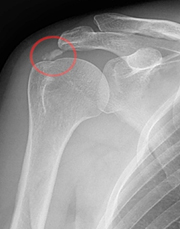
Calcific tendinopathy of the shoulder, often referred to as calcific tendinitis, is characterized by the deposition of calcium hydroxyapatite crystals within the rotator cuff tendons. The condition is a common cause of atraumatic shoulder pain and may range from an incidental radiographic finding to an acute, severely painful inflammatory disorder.
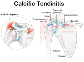
The supraspinatus tendon is most frequently involved, followed by the infraspinatus and subscapularis tendons. It is most commonly seen between 30 and 60 years of age and has a female predominance. Bilateral involvement has been reported in a substantial proportion of patients.
Importantly, calcific tendinopathy should not simply be regarded as a degenerative process, says Dr Ayyappan V Nair, considered as the best shoulder surgeon in India. Current understanding supports a cell-mediated, multiphasic process involving fibrocartilaginous metaplasia, calcium deposition and subsequent spontaneous resorption.
“Calcific tendinopathy is a multifocal, cell-mediated disease.”
— Uhthoff & Loehr, J Am Acad Orthop Surg
This concept is clinically important because the disease can be self-limiting, and the natural history has a major influence on treatment selection.
Why Does Calcific Tendinopathy Occur?
The exact aetiology remains uncertain. Several theories have been proposed over the years, including ischemic, degenerative, metaplastic and genetic mechanisms.
1. Ischemic theory
Early theories proposed that local vascular compromise or microvascular injury could initiate tendon degeneration and subsequent calcification.
McNab suggested that vascular factors may contribute to the development of calcific deposits, although this theory does not completely explain the cell-mediated nature of the disease.
2. Degenerative theory
Degenerative changes in the rotator cuff were initially thought to be responsible for calcium deposition. However, histopathological studies demonstrated that calcific tendinopathy differs from conventional degenerative tendon disease.
3. Multiphasic theory
The multiphasic model described by Uhthoff and colleagues remains one of the most widely accepted explanations.
The process can be broadly divided into:
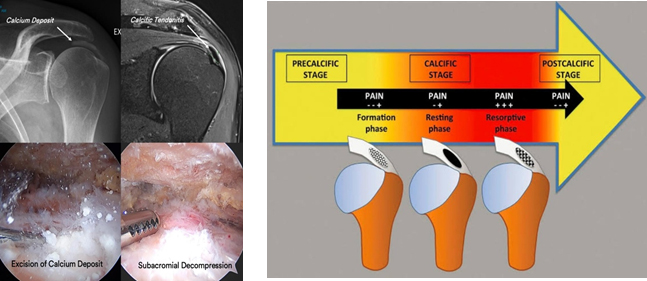
Pre-calcific phase → Calcific phase → Post-calcific/resorptive phase
During the pre-calcific phase, tenocytes undergo fibrocartilaginous metaplasia. Hydroxyapatite crystals subsequently accumulate within the metaplastic tissue.
The calcific phase consists of formative, resting and resorptive stages. During the resorptive phase, macrophages and multinucleated giant cells participate in removal of the calcium deposit, often accompanied by intense inflammation.
This explains an important clinical observation: the most painful episodes frequently occur during the resorptive phase rather than during stable calcification.
4. Metaplastic theory
Another proposed mechanism is transformation of tendon-derived mesenchymal cells into chondrocyte-like or osteogenic cells, resulting in ectopic matrix production and hydroxyapatite deposition.
5. Genetic and molecular mechanisms
Molecular studies have identified altered expression of several genes and proteins involved in mineralization and extracellular matrix regulation, including tissue transglutaminase, osteopontin and bone morphogenetic proteins.
These findings support the concept that calcific tendinopathy is a biologically active tendon disorder rather than simply passive calcium accumulation.
The Stages of Calcific Tendinopathy
Understanding the stage of disease is particularly relevant when deciding treatment.
1. Pre-calcific phase
This stage is characterized by:
- Fibrocartilaginous metaplasia
- Increased proteoglycan production
- Transformation of tenocytes
- Development of fibrocartilaginous tissue
Patients may have minimal or no symptoms during this stage.
2. Calcific phase
Formative phase
Hydroxyapatite crystals accumulate within the fibrocartilaginous tissue. Small deposits gradually coalesce to form larger calcific deposits.
Resting phase
The deposit becomes relatively well defined and is surrounded by fibrocartilaginous tissue. There may be little inflammatory activity, and symptoms may be mild or absent.
Resorptive phase
This is the most inflammatory phase.
The calcium deposit undergoes liquefaction and becomes surrounded by macrophages, multinucleated giant cells and increased vascularity. Calcium may rupture through the tendon into the subacromial bursa, producing an acute inflammatory bursitis.
Patients can present with sudden, severe shoulder pain, marked restriction of movement and night pain, sometimes without any preceding trauma.
3. Post-calcific phase
Following resorption, the tendon undergoes remodeling. Fibroblasts migrate into the defect and new collagen is formed, allowing restoration of the tendon architecture.
Clinical Presentation
The clinical spectrum is broad.
Some patients have an incidental calcific deposit with minimal symptoms, whereas others present with severe disability.
Typical presentations include:
- Atraumatic shoulder pain
- Severe night pain
- Pain during overhead activities
- Painful arc
- Restriction of active and passive movement during acute inflammatory episodes
- Subacromial tenderness
- Chronic shoulder pain in patients with persistent deposits
Acute exacerbation may occur when a deposit enters the resorptive phase and ruptures into the subacromial bursa, producing an intense inflammatory response.
This distinction is important because the radiographic appearance of the deposit alone does not necessarily correlate with symptom severity.
Diagnosis
Diagnosis is usually established by combining the clinical examination with plain radiographs and ultrasound.
Plain Radiographs
Radiographs remain the basic imaging investigation.
Anteroposterior and appropriate rotational views can demonstrate:
- Location of the deposit
- Size
- Density
- Morphology
- Relationship to the greater tuberosity and acromion
Radiographic morphology may also provide information regarding the stage of calcification.
Ultrasound
Ultrasound is particularly useful because it can provide both diagnostic and interventional information.
It allows assessment of:
- Deposit morphology
- Tendon involvement
- Subacromial bursitis
- Rotator cuff integrity
- Doppler vascularity
- Dynamic relationship between the deposit and surrounding structures
Several ultrasound classifications have been proposed.
Farin et al. classified deposits according to acoustic shadowing, while Chiou et al. described patterns including arc-shaped, fragmented, punctuated, nodular and cystic deposits.
From a practical standpoint, the distinction between hard/organized deposits and soft or resorptive deposits can be particularly useful when planning ultrasound-guided lavage.
MRI
MRI is not routinely required to diagnose calcific tendinopathy because calcium is readily identified on radiographs and ultrasound.
However, MRI may be useful when:
- Symptoms are chronic or atypical
- A rotator cuff tear is suspected
- There is concern regarding another intra-articular pathology
- The diagnosis remains uncertain
Calcific deposits typically demonstrate low signal intensity on conventional MRI sequences, with surrounding inflammatory signal potentially visible on fluid-sensitive sequences.
CT
CT is rarely necessary for routine diagnosis but may provide additional information regarding the density and consistency of calcific deposits, particularly when surgical planning is being considered.
Treatment: A Stepwise Approach
The treatment of calcific tendinopathy has changed considerably.
The traditional approach consisted primarily of analgesics, physiotherapy, injections and surgery for persistent symptoms.
Today, treatment is better viewed as a stage-dependent and patient-specific algorithm, incorporating the size and morphology of the deposit, symptom duration, inflammatory phase, functional impairment and response to previous treatment.
Current options include:
- Activity modification and analgesia
- Physiotherapy
- Subacromial corticosteroid injection
- Extracorporeal shockwave therapy
- Ultrasound-guided needling/lavage
- Emerging biological treatments
- Arthroscopic removal of the deposit
1. Conservative Management
For many patients, initial management remains non-operative.
Treatment may include:
- Relative rest and activity modification
- NSAIDs or other analgesics when appropriate
- Physiotherapy
- Restoration of shoulder range of motion
- Rotator cuff and scapular strengthening
- Treatment of associated subacromial bursitis
The self-limiting nature of the disease means that immediate invasive treatment is not necessary in every patient.
What does recent evidence say?
A 2025 network meta-analysis of 33 randomized controlled trials involving 26 treatment strategies found that comprehensive physiotherapy performed particularly well for functional improvement, while shockwave therapy combined with physiotherapy was also among the better-performing approaches.
“Comprehensive PT demonstrates superior efficacy in improving functional outcomes.”
— Yao et al., EFORT Open Reviews, 2025
This supports physiotherapy as an important component of treatment rather than merely an adjunct.
2. Subacromial Corticosteroid Injection
A subacromial corticosteroid injection can be useful in patients with significant inflammatory bursitis, particularly during an acute painful episode.
However, corticosteroid injection primarily addresses the inflammatory component and does not directly remove the calcific deposit.
Therefore, in patients with persistent symptomatic deposits, other interventions may be considered.
3. Extracorporeal Shockwave Therapy
Extracorporeal shockwave therapy (ESWT) is a non-invasive treatment that has become an established option for symptomatic calcific tendinopathy.
Both focused and radial shockwave systems have been studied, with variation in energy levels and treatment protocols.
A 2024 systematic review and meta-analysis including 21 randomized controlled trials found that ESWT was superior to sham treatment for pain and function at medium-term follow-up.
Importantly, the review also suggested that high-energy ESWT may be more effective than low-energy treatment for pain, function and calcific deposit resorption.
“HE-SWT was clinically more effective than LE-SWT.”
— Brindisino et al., Physiotherapy Research International, 2024
The evidence should nevertheless be interpreted cautiously because the certainty of evidence was rated very low in that review.
4. Ultrasound-Guided Needling and Lavage
Ultrasound-guided needling, often called barbotage or ultrasound-guided percutaneous lavage, has become one of the most important minimally invasive treatments for persistent symptomatic calcific deposits.
Under ultrasound guidance, the calcific deposit is punctured and irrigated, allowing fragmentation and aspiration of calcium.
The technique has several potential advantages:
- Minimally invasive
- Direct treatment of the calcific deposit
- Can be performed under local anaesthesia
- Allows simultaneous assessment of the rotator cuff and bursa
- May accelerate radiographic resorption
Current evidence
A 2024 systematic review and meta-analysis of seven randomized trials involving 709 patients found that ultrasound-guided lavage was a reasonable and safe treatment, although differences between lavage and ESWT varied according to follow-up duration and outcome measured. The authors emphasized that certainty of evidence ranged from low to very low.
Another 2024 meta-analysis involving 779 patients from 22 studies found that lavage demonstrated increasing benefit for pain reduction over longer follow-up, while ESWT showed strong improvements in functional scores over time.
“UGL seems to be a reasonable and safe treatment for RCCT.”
— Sconza et al., European Journal of Physical and Rehabilitation Medicine, 2024
Thus, rather than considering lavage and ESWT as competing treatments with one universally superior modality, the choice should be individualized according to deposit characteristics, clinical stage, expertise and patient preference.
5. Platelet-Rich Plasma and Other Biological Treatments
PRP has attracted interest as a biological treatment for rotator cuff disorders.
However, it is important to distinguish evidence for general rotator cuff tendinopathy from evidence specifically supporting PRP for calcific tendinopathy.
A 2025 network meta-analysis of conservative treatments reported a high probability of PRP improving shoulder function, but the evidence base remains heterogeneous and there is currently no universally accepted PRP protocol specifically for calcific deposits.
Therefore, PRP should currently be regarded as an emerging rather than established first-line treatment for calcific tendinopathy.
“No gold standard has been established for managing rotator cuff calcific tendinopathy.”
— Recent systematic review and network meta-analysis, 2025
This is an important distinction when counselling patients regarding newer biological therapies.
6. When Should Surgery Be Considered?
Arthroscopic treatment is generally reserved for patients who remain significantly symptomatic despite an adequate course of non-operative treatment.
Potential indications include:
- Persistent significant pain and functional limitation
- Failure of structured conservative treatment
- Persistent symptomatic calcific deposits despite minimally invasive treatment
- Large or mechanically problematic deposits
- Associated rotator cuff pathology
- Recurrent symptoms affecting quality of life
Historically, a period of approximately 6 months or more of persistent symptoms was often used as a practical threshold before surgery. However, the decision should not be based solely on duration.
The patient’s symptoms, deposit characteristics, response to previous treatment and associated pathology should guide the timing.
Arthroscopic Treatment
Arthroscopy allows direct visualization and removal of the calcific deposit.
The procedure may include:
- Subacromial bursectomy
- Identification and localization of the calcific deposit
- Longitudinal opening of the tendon when required
- Curettage/debridement of calcium
- Assessment of the remaining rotator cuff
- Repair of a significant tendon defect when indicated
The role of routine acromioplasty or subacromial decompression is increasingly questioned.
A 2025 network meta-analysis found that arthroscopic bursectomy and calcific debridement produced similar functional and pain outcomes whether or not subacromial decompression was added.
“ABD alone demonstrated similar clinical effects to ABD + ASD.”
— Yao et al., EFORT Open Reviews, 2025
This suggests that decompression should not necessarily be considered an obligatory component of every calcific deposit excision.
Should the Rotator Cuff Defect Be Repaired?
One of the ongoing controversies in arthroscopic treatment is whether the tendon defect created after calcium removal should routinely be repaired.
A 2025 systematic review and meta-analysis involving 21 studies and 1,172 patients found that both debridement alone and debridement with tendon repair produced substantial improvements in pain and functional scores.
However, the authors reported a higher proportion of complications in the repair group and questioned whether every partial tendon defect requires suturing.
“It is really necessary to repair all partial tears after the calcified deposits removal.”
— González-Martín et al., Revista Española de Cirugía Ortopédica y Traumatología, 2025
In clinical practice, the decision should therefore be individualized according to the size and depth of the residual cuff defect, tendon quality and functional demands of the patient, rather than repairing every defect automatically.
A Practical Treatment Algorithm
Mild symptoms / early presentation
Clinical assessment → X-ray + ultrasound → analgesia/NSAIDs → physiotherapy → observation
↓
Persistent symptoms
Consider:
ESWT
or
Ultrasound-guided needling/lavage
depending on deposit morphology, clinical phase and local expertise.
↓
Persistent disabling symptoms despite non-operative treatment
Reassess with imaging for:
- Persistent calcific deposit
- Rotator cuff pathology
- Subacromial bursitis
- Alternative diagnoses
↓
Refractory symptomatic disease
Arthroscopic calcific deposit removal ± bursectomy ± selective rotator cuff repair
Key Points for Clinical Practice
- Calcific tendinopathy is a cell-mediated, multiphasic disorder, not simply degenerative calcium deposition.
- The supraspinatus is the most commonly involved tendon.
- Symptoms may be particularly severe during the resorptive phase.
- Plain radiographs and ultrasound are usually sufficient for diagnosis.
- Ultrasound is particularly valuable because it can be used for both diagnosis and intervention.
- Physiotherapy remains an important component of non-operative management.
- ESWT is an established non-invasive option, with evidence suggesting a potential advantage of higher-energy protocols.
- Ultrasound-guided needling/lavage is an important minimally invasive option for persistent symptomatic deposits.
- Current evidence does not establish one universally superior non-operative treatment.
- PRP and other biological treatments remain promising but require better disease-specific evidence.
- Arthroscopic removal is appropriate for selected patients with persistent, disabling symptoms despite non-operative treatment.
- Routine subacromial decompression does not appear necessary in every arthroscopic procedure.
- Following deposit removal, rotator cuff repair should be individualized rather than performed routinely for every residual defect.
What Recent Research Tells Us
Evidence Box 1 — 2025 Network Meta-analysis
33 RCTs | 26 treatment strategies
Comprehensive physiotherapy ranked highly for functional improvement, while radial shockwave therapy combined with physiotherapy performed particularly well for pain.
Clinical message: There is no single treatment that should be considered universally superior; treatment should be individualized.
Evidence Box 2 — Ultrasound-Guided Lavage
2024 systematic review | 7 RCTs | 709 patients
Ultrasound-guided lavage appears safe and reasonable, with evidence suggesting potential advantages for long-term pain and calcific deposit resorption, although certainty remains limited.
Clinical message: Lavage is an important minimally invasive option, particularly for appropriately selected deposits.
Evidence Box 3 — Shockwave Therapy
2024 systematic review | 21 RCTs
ESWT was superior to sham treatment for pain and function at medium-term follow-up. High-energy treatment appeared more effective than low-energy treatment for several outcomes.
Clinical message: ESWT remains a valuable non-invasive treatment, particularly when an appropriately selected protocol is used.
Evidence Box 4 — Arthroscopic Treatment
2025 systematic review and meta-analysis | 1,172 patients
Both arthroscopic debridement alone and debridement with repair produced substantial functional improvement. Routine repair of every residual tendon defect may not be necessary.
Clinical message: Tendon repair should be based on the actual structural defect rather than performed automatically.
Conclusion
Calcific tendinopathy of the shoulder is a dynamic biological process with a characteristic natural history. Recognition of the stage of disease, deposit morphology and severity of symptoms is essential for selecting appropriate treatment.
Most patients can be successfully managed without surgery using a combination of rehabilitation, symptom control and appropriately selected minimally invasive interventions. ESWT and ultrasound-guided lavage have emerged as important evidence-supported options for patients with persistent symptoms.
For patients who remain significantly symptomatic despite non-operative treatment, arthroscopic removal of the deposit provides reliable clinical improvement. However, contemporary evidence increasingly supports a selective approach to subacromial decompression and rotator cuff repair, rather than performing these procedures routinely.
The modern management of calcific tendinopathy is therefore not simply about “removing calcium”; it is about understanding the biology of the disease and matching the intervention to the patient, the stage of disease and the characteristics of the deposit.
For enquiries and online appointments, send a message to
www.DrAyyappanVNair.com/contact
For informative videos related to Shoulder problems and their treatment options, Sports Injuries and other orthopedic conditions, visit our YouTube channel Bangalore Shoulder Institute – https://www.youtube.com/@BangaloreShoulderInstitute
Selected Recent References
- Yao Y, Yang G, Jiang S, et al. Treatments for rotator cuff calcific tendinitis: a systematic review and network meta-analysis of randomized-controlled trials. EFORT Open Rev. 2025;10(7):520-533. doi:10.1530/EOR-2024-0078.
- Sconza C, Palloni V, Lorusso D, et al. Ultrasound-guided percutaneous lavage for the treatment of rotator cuff calcific tendinopathy: a systematic review with meta-analysis of randomized controlled trials. Eur J Phys Rehabil Med. 2024;60(6):995-1008. doi:10.23736/S1973-9087.24.08544-7.
- Brindisino F, Marruganti S, Lorusso D, et al. The effectiveness of extracorporeal shock wave therapy for rotator cuff calcific tendinopathy: a systematic review with meta-analysis. Physiother Res Int. 2024;29(3):e2106. doi:10.1002/pri.2106.
- Forogh B, Karami A, Bagherzadeh Cham M. Effect of extracorporeal shock wave therapy and ultrasound-guided percutaneous lavage in reducing the pain of rotator cuff calcific tendinopathy: an updated systematic review and meta-analysis. J Orthop. 2024;56:151-160. doi:10.1016/j.jor.2024.05.023.
- González-Martín D, Garrido-Miguel M, de Cabo G, et al. Rotator cuff debridement compared with rotator cuff repair in arthroscopic treatment of calcifying tendinitis of the shoulder: a systematic review and meta-analysis. Rev Esp Cir Ortop Traumatol. 2025;69(1):91-103. doi:10.1016/j.recot.2023.08.015.
- Romano AM, et al. Efficacy of conservative approaches on pain relief and function in patients with rotator cuff calcific tendinopathy: which is the best option? A systematic review and network meta-analysis. Orthop Surg. 2025. doi:10.1111/os.70175.