Journal of ISAKOS – Aug 2025
LINK: https://www.jisakos.com/article/S2059-7754(25)00613-3/fulltext4
The posterolateral corner (PLC) of the knee is crucial for maintaining varus and rotational stability, and its injuries, often associated with cruciate ligament tears, pose major reconstructive challenges. Grade III PLC injuries, defined by significant varus opening and external rotation on stress testing, require surgical reconstruction. Traditionally, LaPrade’s anatomic technique using Achilles tendon allograft is considered the gold standard, but limitations in graft availability have prompted autograft-based alternatives.
This article presents a single-stage approach combining anatomic PLC reconstruction with a modified LaPrade technique and simultaneous posterior cruciate ligament (PCL) reconstruction, performed in a patient with a tibial intramedullary nail in situ after a high-energy fracture. The technique utilizes a Y-shaped peroneus longus autograft for PLC reconstruction and hamstring tendons for PCL reconstruction. The graft passage and fixation sequence are reversed from the classic LaPrade method, allowing independent tensioning of each structure (lateral collateral ligament, popliteus, and popliteofibular ligament) while preserving biomechanics.
Clinical outcomes showed significant functional improvement, restoration of knee stability, and recovery of range of motion, with no early graft failure. This technique offers a cost-effective, autograft-based solution for complex multiligament injuries, particularly when allografts are unavailable, and demonstrates promising results in restoring stability and function in knees with combined PLC and PCL injuries.
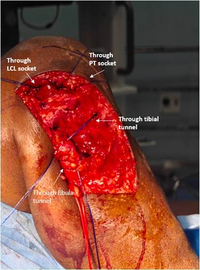
Single-stage anatomic posterolateral corner reconstruction with modified LaPrade technique, combined with arthroscopic posterior cruciate ligament reconstruction (tibia nail in situ)
- Patient positioning:
Supine with a high tourniquet; both knees flexed to 90◦ using a foot support and lateral thigh post.
- Y-graft preparation:
Peroneus longus (≥250 mm), semitendinosus, and gracilis tendons harvested from the contralateral limb. Peroneus longus used for posterolateral corner and hamstrings for posterior cruciate ligament reconstruction (Fig. 1).
- Surgical approach and nerve neurolysis:
Perform standard exposure and common peroneal nerve neurolysis (Fig. 2A).
- Soft tissue windows:
○ Posterior: Via peroneal fascia for tibial popliteal sulcus (Fig. 2B)
○ Middle: Between the iliotibial band and biceps femoris to expose the lateral collateral ligament (Fig. 2C) ○ Anterior: Through iliotibial band to identify lateral epicondyle and femoral-popliteal sulcus (Fig. 2D)
• Tunnel Preparation:
○ Femoral tunnels: Lateral collateral ligament (LCL) and popliteus sockets created anatomically with a minimum 18.5 mm bone bridge; reamed with 7 × 25 mm reamers (Fig. 3).
○ Fibular tunnel: Drilled 28 mm distal to the styloid tip, angled posterosuperomedially (Fig. 4).
○ Tibial tunnel: Retrodrilled from posterior cortex using an 8 mm FlipCutter (Arthrex, Naples, FL) guided by a posterolateral corner (PCL) jig (Fig. 5).
- Suture loop passage:
Loops passed through all tunnels for graft shuttling (Fig. 5).
- Graft passage & fixation:
○ The adjustable loop is shuttled from posterior to anterior until the loop button sits flat on the tibia (Fig. 7A).
○ Y-graft long limb passed through the fibular tunnel (posterior to anterior) (Fig. 7B).
○ Short limb fixed in femoral popliteus socket with a 7 × 23 mm screw at 90◦ knee flexion (Fig. 7C).
○ Long limb fixed in fibula with a 6 × 23 mm screw, recreating the popliteofibular ligament (Fig. 8A).
○ The remaining limb is fixed in femoral LCL socket with a 7 × 23 mm screw at 30◦ flexion to recreate the lateral collateral ligament (Fig. 8B and C).
• Arthroscopic posterior cruciate ligament reconstruction: Standard arthroscopic PCL reconstruction using hamstring graft (Fig. 9).
Introduction
Outline of the clinical problem
The three main structures constituting the posterolateral corner (PLC) are lateral collateral ligament (LCL), popliteus (PT), and popliteofibular ligament (PFL). Grade III injuries, defined clinically using the Modified Hughston criteria, result in varus and rotational instability throughout the range of motion, necessitating surgical reconstruction of the affected structures [1]. The PLC injuries rarely occur in isolation, with a notably high incidence of associated anterior cruciate ligament (ACL) or posterior cruciate ligament (PCL) injuries, ranging from 43% to 80% [2]. Identifying these concomitant injuries is crucial not only for restoring biomechanics but also for reducing stress on other reconstructed ligaments. The mechanism of injury involves twisting of the knee, hyperextension, or direct varus stress. Chronic posterolateral instability can lead to varus thrust gait, causing considerable changes in medial and patellofemoral compartments, resulting in meniscal injuries and early osteoarthritis [3]. LaPrade described the anatomical reconstruction of PLC using a split Achilles tendon allograft. Due to the limited availability and high cost of allografts in many countries, our technique offers an alternative approach. In our technique, we use the modified LaPrade technique for anatomical reconstruction of PLC structures using a single peroneus longus tendon autograft. Subsequently, we performed arthroscopic reconstruction of PCL using hamstring tendons as a single-staged surgery. A similar technique has been described by Tapasvi et al. but for an isolated PLC injury [4] (see Fig. 6)
Our patient is a 30-year-old male, who had a road traffic accident and sustained Type IIIb open fracture shaft of tibia with peroneal nerve injury. He underwent intramedullary nailing and fasciocutaneous flap reconstruction. Six months later, he presented with varus laxity and rotational instability (Figs. 10 and 11). Clinical examination and magnetic resonance imaging (MRI) revealed a complete tear of PLC and PCL (Figs. 12 and 13).
The Technique
Surgical indications/contraindications
PLC injuries are clinically classified into three grades according to the modified Hughston classification: Grade I (<5 mm lateral opening on varus stress in slight flexion, no rotational laxity on dial test), Grade II (6–10 mm opening with 5–10◦ difference in external rotation), and Grade III (>10 mm opening with >10◦ external rotation difference on dial test). PLC reconstruction is indicated in Grade III injuries, which reflect complete structural disruption and are confirmed by using MRI [5]. PCL reconstruction is indicated in isolated complete tears and associated multiligament injuries. Contraindications include active infection, uncontrolled comorbidities, arthrofibrosis, chronic genu varus malalignment greater than 5◦, combined tibial and peroneal nerve injuries, and advanced knee arthritis [6].
Treatment options
Grade I and II PLC injuries can be treated conservatively. Nonoperative management of Grade III PLC injuries not only displays persistent lateral instability but also commonly includes multidirectional knee instability. Therefore, surgical management is recommended to restore knee stability in Grade III PLC injuries [7].
The patients who undergone PLC repair have been found to have more reoperation rates due to failure when compared to the reconstruction group. Hence, reconstruction is the treatment of choice for Grade III PLC injuries [7]. Persistent posterolateral instability with Grade III injuries has been found to have an increased risk of meniscal injuries and early osteoarthritis. They also imply more force on ACL and PCL grafts in the setting of multiligamentous knee injuries, causing graft failure [8].
Our patient exhibited a severe varus thrust gait; therefore, a biplanar medial opening wedge osteotomy was another option, allowing for an
Fig. 1. Y-configuration peroneus longus graft preparation for PLC reconstruction: At least 250 mm long graft is required. (A) Mark 1 made at 25 mm from distal end, mark 2 at 45 mm from mark 1, and mark 3 at 25 mm from mark 2. The distal 25 mm upto mark 1 is whipstitched with No.2 FiberWire. (B) The adjustable loop thread is looped around mark 3 and the length of 25 mm looped around the thread is sutured to prevent slippage. (C) The final Y-configuration has a distal 25 mm to be inserted into the femoral popliteal socket, the short limb of 45 mm to recreate popliteus, and the longer limb of Y construct to reconstruct PFL and LCL. The looped graft of 25 mm inserted into the tibial tunnel for suspensory fixation, for PT, and PFL. PT = popliteus tendon; PFL = popliteofibular ligament; LCL = lateral collateral ligament.
’ lateral side. The popliteal femoral socket is created at the anterior fifth of the popliteal sulcus using a 7 mm reamer up to a depth of 25 mm. The LCL socket is created at a point 1.4 mm proximal and 3.1 mm posterior to the lateral epicondyle using a 7 mm reamer up to a depth of 25 mm. The distance between the two sockets is about 18.5 mm, which is the normal anatomical distance between PT and LCL. PT = popliteus tendon; LCL = lateral collateral ligament.
Fig. 4. Fibular tunnel placement: An intraoperative image of the right knee’s lateral side. The fibula tunnel is created at a point 28 mm distal from the styloid tip. The tunnel is directed from the anterolateral aspect (corresponding to the Fibular collateral ligament attachment) to the posteromedial aspect (corresponding to the PFL attachment), angling posterosuperomedially. The tunnel is drilled using a 6 mm reamer to prevent fracture. PFL = popliteofibular ligament.
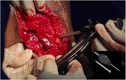
Fig. 5. Tibial tunnel placement: An intraoperative image of the right knee’s lateral side. A PCL femur jig is placed at the tibial popliteal sulcus posteriorly which is 1 cm distal from the joint line. Entry is made at a point just medial and distal to Gerdy’s tubercle. Retrodrilling of tibia performed using an 8 mm Flipcutter (shown in accessory window) upto a depth of 28 mm. PCL = posterior cruciate ligament.
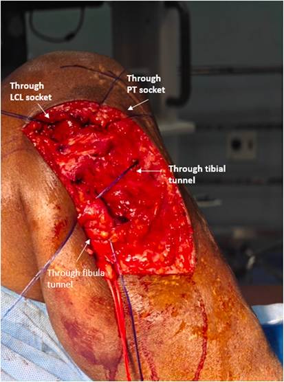
Fig. 6. An intraoperative image of the right knee’s lateral side. Suture loops are passed through each tunnel after the tunnel placement for graft passage. PT = popliteus tendon; LCL = lateral collateral ligament.
increase in the posterior slope and lateral translation of the mechanical axis to address posterior and posterolateral instability [9]. If necessary, PCL and PLC reconstruction could be considered as a second-stage procedure. However, in this case, osteotomy was not feasible as the intramedullary tibial nail obstructs the bone cuts and the placement of osteotomy fixation plates. Moreover, removing the nail was not advisable as the fracture site had not yet fully united. As a result, staged correction with implant removal and osteotomy was deferred, and a single-stage reconstruction of the PCL and PLC was planned.
Outcome of the technique
Anatomical reconstruction is the gold standard for Grade III PLC injuries. Laprade et al. demonstrated the reconstruction at the native attachment of LCL, PL, and PFL, thereby restoring the varus and rotational stability of the knee [10]. Studies have shown excellent improvement in postoperative scores following reconstruction. Geeslin et al. on studying the anatomical reconstruction of PLC demonstrated significant improvement in IKDC (International Knee Documentation Committee) scores, the side-to-side difference in varus stress radiograph, and mean Cincinnati scores [5].
There has been no conflict of interest among surgeons regarding reconstruction as an option for chronic injuries. However, in acute injuries, some advocate repair over reconstruction. Levy et al. found an overall failure rate of 40% in the repair group compared to 6% in the reconstruction group when evaluating patients with PLC tears [7]. In our patient, following graft fixation, time-zero stability was confirmed intraoperatively, with the dial test demonstrating symmetrical external rotation at 30◦ and 90◦, and the varus stress test showing less than 3 mm of opening with a firm endpoint. At 6-month follow-up, the patient demonstrated significant improvement, with the Lysholm score increasing from 19 (post tibial nailing) to 74, full range of motion (0–120◦) without clinical instability, and with resolution of varus. The IKDC subjective score is 78.2%. The concomitant peroneal nerve injury was managed conservatively with splinting, physiotherapy, and serial clinical follow-up. At the latest follow-up, there were signs of neurological improvement.
Complications
The complications of PLC and PCL reconstruction include persistent instability, popliteal artery injury, common peroneal injury, arthrofibrosis, compartment syndrome due to fluid extravasation, etc [3,10]. However, addressing concomitant injuries, proficiency with surgical dissection, and proper anatomical placement of tunnels reduces these complications.
Conclusion and future perspectives
This surgical approach demands expertise in dissection and a deep understanding of anatomical structures. Utilizing advanced techniques and structured rehabilitation, our technique of anatomic reconstruction effectively restores knee stability and function in multiligament injuries.
Future advancements in surgical techniques, biologics, rehabilitation, and technology will continue to refine PCL and PLC reconstruction, leading to improved patient outcomes, faster recovery, and better long- term knee function. Ongoing research will determine the optimal grafts, fixation methods, and treatment combinations for different patient populations.
Fig. 7. Popliteus tendon reconstruction: An intraoperative image of the right knee’s lateral side. (A) The adjustable loop is shuttled from posterior to anterior until the button sits flat on the tibia. The long limb is shuttled through fibula from posterior to anterior. (B) The short limb of the graft is passed deep into the IT band and inserted into the femoral popliteal socket. (C) The fixation is performed by a 7 × 23 mm BioComposite screw keeping the knee in 90◦ of flexion and gentle valgus. PT = popliteus tendon; IT band = iliotibial band.
Fig. 8. Popliteofibular and lateral collateral ligament reconstruction: An intraoperative image of the right knee’s lateral side. (A) The longer limb that is shuttled through the fibula is held tight and a 6 × 23 mm BioComposite screw inserted from anterior to posterior, to reconstruct popliteofibular ligament. (B) The remaining longer limb is shuttled deep to ITB and superficial to popliteus tendon and inserted to femoral LCL socket. (C) Fixation performed with a 7 × 23 mm BioComposite screw keeping the knee in 30◦ of flexion to reconstruct the lateral collateral ligament. ITB = iliotibial band; LCL = lateral collateral ligament; PT = popliteus tendon.
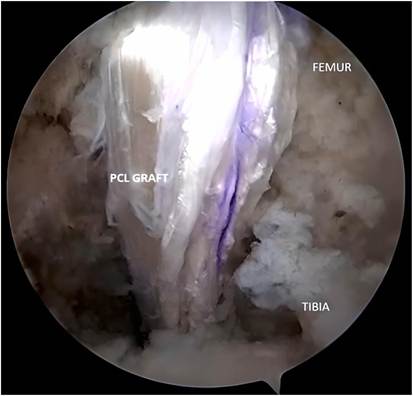
Fig. 9. The patient in supine position. An arthroscopic view of the right knee showing posterior cruciate ligament graft.
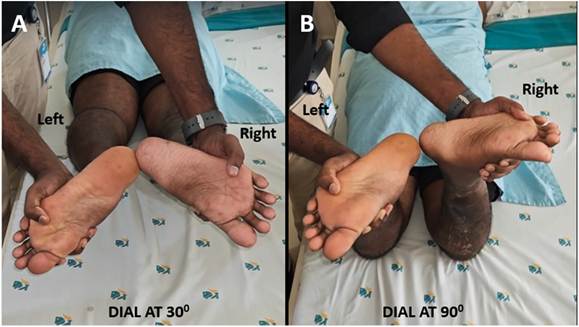
Fig. 10. Performing dial test at (A) 30◦ and (B) 90◦ of knee flexion. There is more than 10◦ of external rotation of the injured right knee when compared to the left side. This is considered to be a positive dial test and indicated injury to both PLC and PCL. PCL = posterior cruciate ligament; PLC = posterolateral corner.
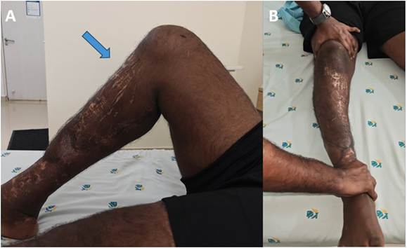
Fig. 11. (A): Posterior sag sign positive. The tibia of the affected side sags posteriorly relative to the femur which indicates PCL tear. (B) Varus stress test positive. The varus stress test at full extension shows excessive lateral gapping with no end point, suggestive of LCL, PLC, and PCL injury. LCL = lateral collateral ligament; PCL = posterior cruciate ligament; PLC = posterolateral corner.
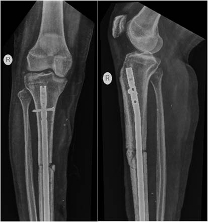
Fig. 12. Preoperative radiograph anteroposterior and lateral view of the right knee showing uniting fracture tibia with nail in situ.
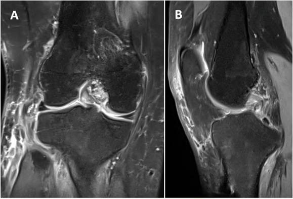
Fig. 13. Proton Density Fat Saturation (PDFS) MRI (A) coronal and (B) sagittal view of the right knee showing complete tear of PCL and PLC structures. MRI = magnetic resonance imaging; PCL = posterior cruciate ligament; PLC = posterolateral corner.
References
- Sajjadi MM, Behroozi A, Matini SA. A modified LaPrade technique in posterolateral corner reconstruction of the knee. Arthrosc Tech 2022;11(3):e413–7. https://doi. org/10.1016/j.eats.2021.11.010. Published 2022 Feb 18.
- Yoon HK, Park SH, Oh HC, Ha JW, Choi H. Combined PCL and PLC reconstruction improves residual laxity in PCL injury patients with posterolateral knee laxity less than grade III. Yonsei Med J 2023;64(5):313–9. https://doi.org/10.3349/ ymj.2022.0487.
- Serra Cruz R, Mitchell JJ, Dean CS, Chahla J, Moatshe G, LaPrade RF. Anatomic posterolateral corner reconstruction. Arthrosc Tech 2016;5(3):e563–72. https:// doi.org/10.1016/j.eats.2016.02.006. Published 2016 Jun 6.
- Tapasvi SR, Shekhar A, Patil SS. Anatomic posterolateral corner reconstruction with autogenous peroneus longus Y graft construct. Arthrosc Tech 2019;8(12):
- e1501–9. https://doi.org/10.1016/j.eats.2019.07.024. Published 2019 Nov 11.
- Geeslin AG, LaPrade RF. Outcomes of treatment of acute grade-III isolated and combined posterolateral knee injuries: a prospective case series and surgical technique. J Bone Joint Surg Am 2011;93(18):1672–83. https://doi.org/10.2106/ JBJS.J.01639.
- Rodriguez AN, Banks E, Monson J, LaPrade RF. Surgical principles for lateral collateral and posterolateral knee injuries. Operat Tech Sports Med 2022 Jun 1;30 (2):150912. https://doi.org/10.1016/j.otsm.2022.150912.
- Levy BA, Dajani KA, Morgan JA, Shah JP, Dahm DL, Stuart MJ. Repair versus reconstruction of the fibular collateral ligament and posterolateral corner in the multiligament-injured knee. Am J Sports Med 2010;38(4):804–9. https://doi.org/ 10.1177/0363546509352459.
- LaPrade RF, Muench C, Wentorf F, Lewis JL. The effect of injury to the posterolateral structures of the knee on force in a posterior cruciate ligament graft: a biomechanical study. Am J Sports Med 2002;30(2):233–8. https://doi.org/ 10.1177/03635465020300021501.
- Savarese E, Bisicchia S, Romeo R, Amendola A. Role of high tibial osteotomy in chronic injuries of posterior cruciate ligament and posterolateral corner. J Orthop Traumatol 2011;12(1):1–17. https://doi.org/10.1007/s10195-010-0120-0.
- LaPrade RF, Johansen S, Agel J, Risberg MA, Moksnes H, Engebretsen L. Outcomes of an anatomic posterolateral knee reconstruction. J Bone Joint Surg Am 2010;92 (1):16–22. https://doi.org/10.2106/JBJS.I.00474.