Written by: Editorial Team of Dr Ayyappan V Nair
Medically Reviewed by: Dr. Ayyappan V. Nair
Senior Consultant – Shoulder Surgery, Arthroscopy and Sports Trauma
Manipal Hospitals, Bangalore
Last Reviewed: 20th September 2026
THE KINETIC CHAIN
- The scapula acts as the vital link between
- Core/trunk (force generation) and
- Arm (force delivery)
- Dyskinesia breaks this chain, leading to energy leaks and secondary shoulder pathologies (e.g., impingement, labral tears)
DYNAMIC ROLE OF SCAPULA
- Provides a stable base for the glenohumeral (GH) joint.
- Elevates the acromion to prevent impingement during abduction/flexion.
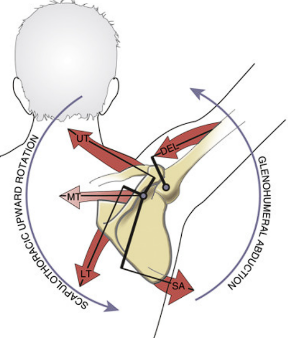
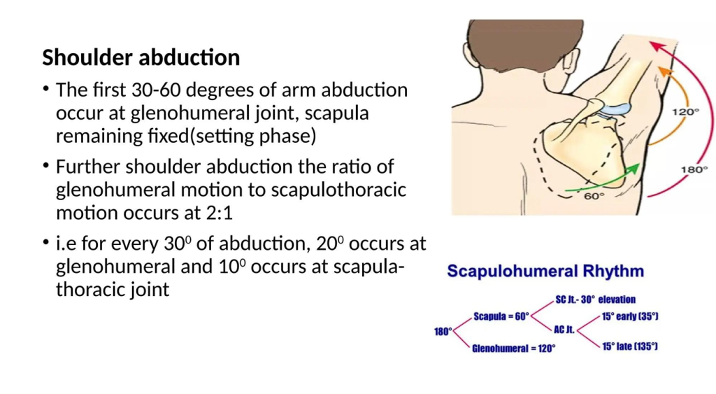
SCAPULO THORACIC KINEMATICS DURING SHOULDER ABDUCTION
- Upward Rotation
- Posterior Tilt
- External Rotation
- Driven by the force couple of the upper trapezius, lower trapezius, and serratus anterior
Movements:
- Upward/downward rotation around a horizontal axis perpendicular to the plane of the scapula
- Internal/external rotation around vertical axis through the plane of the scapula
- Anterior/posterior tilt around a horizontal axis in the plane of the scapula
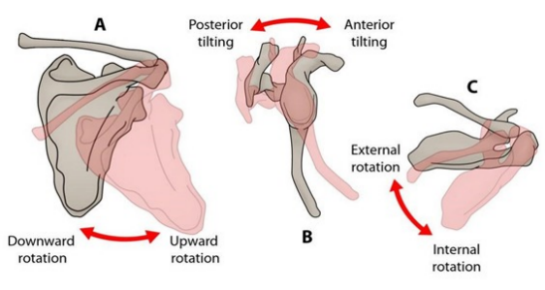
Coupling Movements of Scapula
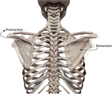
- Retraction : external rotation, posterior tilt, upward rotation and medial translation
- Protraction : internal rotation, anterior tilt, downward rotation and lateral translation
- Shrug : upward translation, anterior tilt and internal rotation
- Coupling movements maintain glenoid as congruent socket, maximizing concavity compression and kinematics
- Scapula stabilized in retraction posture during arm elevation to achieve maximal activation of all muscles originating on the scapula
ETIOLOGY OF SCAPULA DYSKINESIA
- Muscle & Nerve:
- Muscle Imbalance: Weakness in the serratus anterior and lower trapezius; tightness in the pectoralis minor and upper trapezius.
- Neurologic Deficits: Long thoracic nerve palsy (serratus anterior), spinal accessory nerve palsy (trapezius), or dorsal scapular nerve involvement.
- Intra-articular Pathology
- The scapula alters its mechanics to compensate for the failing GH joint in Instability, SLAP tears, or rotator cuff disease.
- Bony/Structural Causes:
- Clavicle fractures (malunion leading to shortened strut).
- Thoracic kyphosis or postural deficits.
SICK SCAPULA SYNDROME
- Coined by Burkhart et al. to describe the pathological state of the scapula in the overhead athlete.
- S: Scapular malposition (typically protracted, anteriorly tilted, and inferiorly translated).
- I: Inferior medial border prominence.
- C: Coracoid pain and malposition (due to chronic pectoralis minor tension).
- K: dysKinesis of scapular movement.
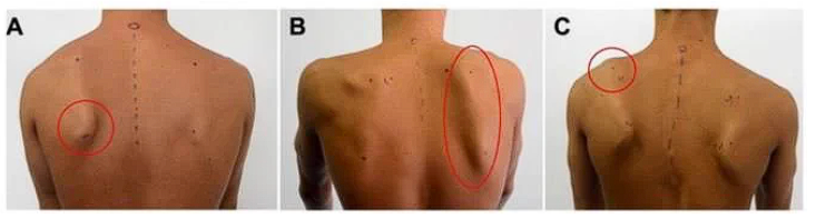
KIBLER CLASSIFICATION
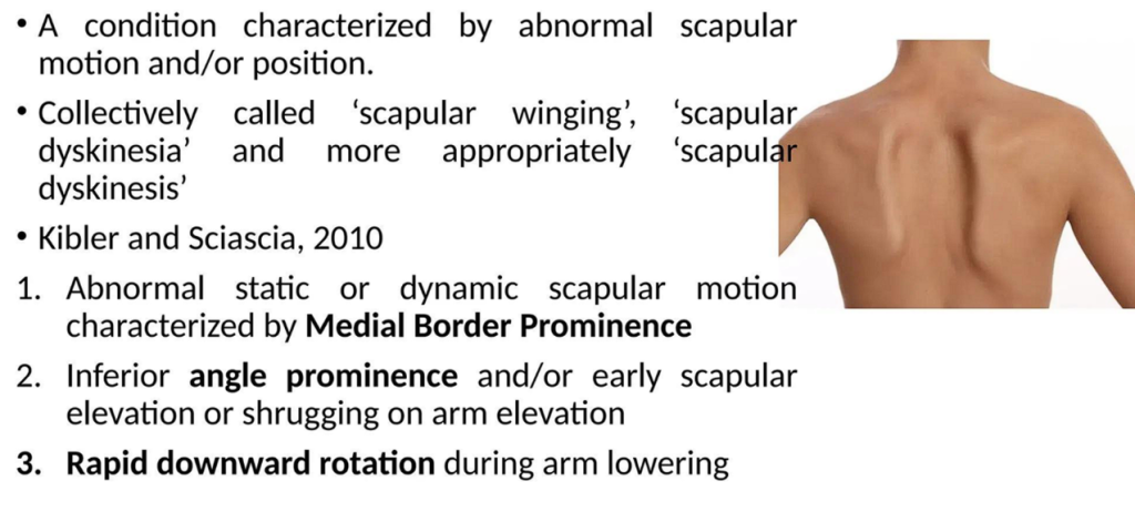
- Type I: Inferior medial border prominence (associated with anterior tilt; common in rotator cuff dysfunction).
- Type II: Entire medial border prominence (winging; indicates internal rotation of the scapula, often serratus anterior weakness).
- Type III: Superior medial border prominence (associated with early, excessive scapular elevation; often involves upper trapezius overactivity).
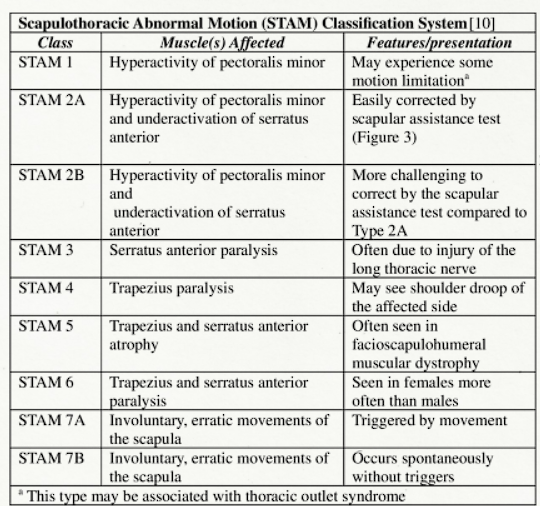
CLINICAL EVALUATION
- Visual Inspection: Evaluate resting posture from behind, observing for asymmetry.
- Dynamic Assessment:
- Observe active elevation/descent in forward flexion.
- Look for a “catch” or dysrhythmia, particularly during the eccentric lowering phase.
- Tenderness at Coracoid tip
- Due to Pectoralis Minor tightness
- Not to be confused with bicipital groove tenderness
EVALUATION
- Radiographs: True AP, Scapular Y, Axillary views (to rule out bony abnormalities, tumors, or fracture malunions).
- MRI: Essential for ruling out causes of dyskinesia (SLAP lesions, rotator cuff tears, Bankart lesions).
Look for signs of paralytic muscle atrophy (denervation changes).
- EMG / Nerve Conduction Studies: Indicated if persistent winging is present or if a specific nerve palsy (Long Thoracic or Spinal Accessory) is suspected.
REHABILITATION – THE MAINSTAY OF TREATMENT
- The vast majority of scapular dyskinesia is treated non-operatively through targeted rehabilitation.
- Phase 1 (Acute/Symptom Alteration): Pain control, soft tissue mobilization (pec minor release), and restoring normal resting posture.
- Phase 2 (Dynamic Stability – Closed Chain): Wall push-ups, weight-bearing shifts. Promotes co-contraction and proprioception without high stress on the GH joint.
- Phase 3 (Dynamic Stability – Open Chain): Targeted strengthening of the lower trapezius and serratus anterior; integrating trunk and hip extensions.
- Phase 4 (Sport-Specific): Plyometrics, throwing programs, focusing on the entire kinetic chain from the legs/hips through the core to the arm.
COMPONENTS OF REHAB
- RELEASING THE THIGHT MUSCLE – Pectoralis minor
- STRENGTHENING THE WEAK MUSCLES – SA , Trapezius
SURGICAL CONSIDERATIONS
- Surgery is rarely indicated for dyskinesia itself; rather, it targets the underlying pathology.
- Addressing Intra-articular Drivers: Arthroscopic SLAP repair, labral stabilization, or rotator cuff repair. Post-operatively, normal kinematics often return as pain and mechanical blocks are removed.
- Addressing Contractures: Surgical release of a severely contracted pectoralis minor (rare, usually conservative).
For informative videos related to Shoulder problems and their treatment options, Sports Injuries and other orthopedic conditions, visit our YouTube channel Bangalore Shoulder Institute – https://www.youtube.com/@BangaloreShoulderInstitute