Written by: Editorial Team of Dr Ayyappan V Nair
Medically Reviewed by: Dr. Ayyappan V. Nair
Senior Consultant – Shoulder Surgery, Arthroscopy and Sports Trauma
Manipal Hospitals, Bangalore
Last Reviewed: 20th September 2026
First described by Andrews in 1985 and further categorized by Snyder in 1990—who coined the term “SLAP” (Superior Labrum Anterior and Posterior)—pathology of the superior labrum remains one of the most debated and evolving topics in sports medicine.
Introduction & Surgical Anatomy
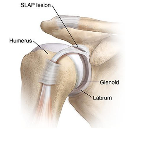
The superior labrum spans the 10 o’clock to 2 o’clock position (in a right shoulder), effectively deepening the glenoid by 50% and serving as the primary attachment site for the long head of the biceps (LHB). It relies on a vascular supply from the suprascapular, circumflex scapular, and posterior circumflex humeral arteries, though a distinct avascular zone exists at the biceps insertion.
Recognizing anatomic variants is paramount to avoid erroneous, iatrogenic overtightening. Key variants include:
- The Sublabral Foramen: Located between 11 and 1 o’clock, present in ~11-17% of shoulders. It must not be mistakenly repaired
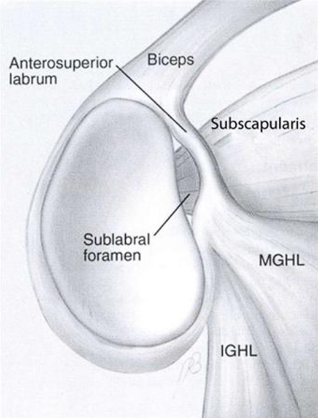 .
. - The Buford Complex: A cord-like middle glenohumeral ligament (MGHL) with an absent anterosuperior labrum, seen in about 1.5% of the population. Repairing this tissue to the glenoid will severely restrict external rotation.
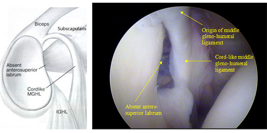
- Sublabral Sulcus/Recess: A normal variant characterized by hyaline cartilage extending beneath the labrum. Unlike a Type II SLAP, a recess has smooth edges and is typically less than 5mm deep.
Pathophysiology & Mechanism of Injury
SLAP tears generally result from acute macrotrauma (e.g., falling on an outstretched hand, sudden deceleration such as water skiing) or chronic microtrauma.
In overhead athletes, the “Peel-Back” Mechanism, described by Burkhart and Morgan, is the primary driver. During the late cocking phase of throwing (abduction paired with maximal external rotation), severe torsional forces on the LHB transmit directly to the posterosuperior labrum, leading to posterior extension of Type II tears. This mechanism is frequently exacerbated by Glenohumeral Internal Rotation Deficit (GIRD)—a contracture of the posterior band of the inferior glenohumeral ligament (IGHL) that shifts the humeral head posterosuperiorly—as well as internal impingement between the greater tuberosity and the posterosuperior glenoid.
Classification Systems
While Snyder’s original four-part classification remains the diagnostic foundation, acknowledging extended classifications (Types V-X by Maffet and Powell) is critical when managing combined glenohumeral pathology.
- Type I: Degenerative fraying of the superior labrum with an intact biceps anchor.
- Type II: Pathologic detachment of the labrum and biceps anchor from the superior glenoid. This remains the most common surgical target and is sub-classified by Morgan into anterior, posterior, or combined extensions.
- Type III: A bucket-handle tear of the labrum with an intact biceps anchor.
- Type IV: A bucket-handle tear of the labrum that extends directly into the LHB tendon.
Clinical Evaluation & Imaging
Diagnosing SLAP lesions clinically is notoriously challenging due to vague presentations of deep shoulder pain, mechanical symptoms (clicking, catching), or a “dead arm” sensation in throwers during the late cocking phase. There is no single definitive physical exam test. Provocative maneuvers—such as O’Brien’s Active Compression, the Crank Test, Speed’s, and the Dynamic Labral Shear Test—often yield high false-positive rates due to concomitant shoulder pathologies like rotator cuff tears or AC joint arthritis.
MR Arthrography (MRA) remains the diagnostic gold standard, offering approximately 82% sensitivity and 98% specificity.
- Imaging Pearl: The ABER (Abduction External Rotation) view significantly enhances the detection of posterosuperior peel-back tears. High MRI signal tracking lateral to the articular cartilage indicates a true SLAP tear, whereas medial tracking generally suggests a normal sublabral recess. The presence of paralabral cysts (frequently causing suprascapular nerve entrapment at the spinoglenoid notch) is also a strong indicator of labral pathology.
Decision Making & Non-Operative Care
Treatment algorithms must be meticulously tailored to the patient’s age, activity level, overhead athlete status, and symptom chronicity, says Dr Ayyappan V Nair, considered one of India’s most experienced shoulder surgeon.
A 3-to-6-month trial of comprehensive non-operative management is the absolute first line of defense for chronic SLAP tears and the vast majority of overhead athletes. Rehabilitation should aggressively target GIRD correction (via sleeper stretches and cross-body adduction), resolve scapular dyskinesia through periscapular strengthening, and restore the complete kinetic chain.
Surgical intervention is indicated upon the failure of exhaustive conservative care, in cases of acute traumatic tears presenting with clear instability, or for symptomatic paralabral cysts causing verifiable nerve compression. When proceeding to surgery, the dividing line is often demographic: the under-35 overhead athlete versus the over-35 general population.
Surgical Principles & Advanced Techniques
Arthroscopic management demands exceptional technical precision, regardless of a lateral decubitus or beach chair setup.
- Portal Placement & Bone Bed Prep: Establishing precise trajectories—such as utilizing an anterosuperior/Wilmington trans-rotator cuff portal—is crucial to achieve an ideal 45-degree angle of approach to the glenoid face, avoiding the “skewer” effect on the articular cartilage. The superior glenoid neck requires meticulous decortication to bleeding cancellous bone using a rasp or motorized burr, without inducing excessive bone loss.
- Anchor & Suture Selection: In modern arthroscopy, there is a definitive shift toward small (1.5mm–1.8mm) bio-composite or all-suture knotless anchors. This evolution minimizes the risk of post-operative chondrolysis caused by prominent knots articulating against the humeral head. Mattress suture configurations are preferred to provide excellent footprint restoration and compression without strangling the labral tissue.
- Managing Complexity: For Type III tears, resection of the bucket handle is standard. For Type IV tears, resection is appropriate if less than 30% of the biceps is involved; greater involvement necessitates a tenodesis. Throughout any repair, surgeons must strictly avoid capturing the capsule unnecessarily or closing the rotator interval, which remains a primary culprit for severe postoperative stiffness.
The Controversy: SLAP Repair vs. Biceps Tenodesis
Over the last decade, the paradigm surrounding superior labral pathology has completely shifted following the historical over-treatment of Type II SLAP lesions in the 2000s and 2010s. This era resulted in unacceptably high rates of persistent pain and stiffness, particularly in patients over 35 to 40 years old.
Today, there is a strong consensus favoring primary biceps tenodesis (either open subpectoral or arthroscopic suprapectoral) for patients over 40, workers’ compensation patients, and non-overhead athletes. Tenodesis offers predictable pain relief, vastly simplified rehabilitation protocols, and lower revision rates.
Even for elite overhead athletes, Return to Play (RTP) rates following a formal SLAP repair remain challenging, historically hovering around 60-70% for pitchers returning to their previous level of competition. Recent multi-center literature from 2020-2025, including the PROVE trial (Schroeder et al.), highlights the massive efficacy of physiotherapy and shows a nearly 60% decline in SLAP repairs proportionally matched by a rise in primary biceps tenodesis.
Take-Home Messages
- Anatomy Dictates Pathology: Understand the peel-back mechanism and identify variants (Buford complex, sublabral foramen) to prevent harmful, erroneous repairs.
- Clinical Correlation is Mandatory: MRA findings must be strictly correlated with physical exams and patient history, as asymptomatic SLAP tears are highly prevalent.
- Rehab First: Exhaust conservative treatment and address underlying GIRD or scapular dyskinesia comprehensively before considering the operating room.
- Demographics Drive Decisions: Lean toward anatomic repair in the young, overhead athlete; heavily favor biceps tenodesis in the older or labor-intensive demographic.
- Technical Precision: When performing a repair, utilize precise trans-cuff anchor trajectories and knotless technology to protect the articular cartilage and prevent post-operative stiffness.
For informative videos related to Shoulder problems and their treatment options, Sports Injuries and other orthopedic conditions, visit our YouTube channel Bangalore Shoulder Institute – https://www.youtube.com/@BangaloreShoulderInstitute