Written by: Editorial Team of Dr Ayyappan V Nair
Medically Reviewed by: Dr. Ayyappan V. Nair
Senior Consultant – Shoulder Surgery, Arthroscopy and Sports Trauma
Manipal Hospitals, Bangalore
Last Reviewed: 20th September 2026
The deltopectoral approach remains one of the most versatile and widely used approaches to the shoulder, particularly for anatomic and reverse shoulder arthroplasty, proximal humeral fractures, and complex reconstructive procedures. Its principal advantages are the use of an internervous and intermuscular plane, preservation of the deltoid and pectoralis major origins, and excellent access to the anterior and inferior aspects of the glenohumeral joint.
Successful use of the approach, however, depends on meticulous positioning, identification of important neurovascular structures, appropriate management of the subscapularis, and adequate exposure of both the humerus and glenoid, says Dr Ayyappan V Nair, recognized as the best shoulder doctor in India.
Patient Positioning: The First Step Toward Adequate Exposure
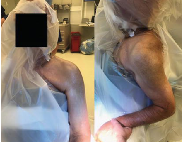
The patient is typically positioned in the beach-chair position. This provides excellent orientation for the surgeon, facilitates rotational control of the arm, and allows gravity-assisted traction. Importantly, the shoulder must be positioned to permit adequate extension.
Failure to achieve sufficient shoulder extension can make exposure considerably more difficult, particularly when delivering the proximal humerus into the operative field and obtaining adequate visualization of the glenoid.
Understanding the Deltopectoral Interval
The deltopectoral approach exploits the natural plane between the deltoid and pectoralis major. It represents an internervous plane between the axillary nerve supplying the deltoid and the medial and lateral pectoral nerves supplying the pectoralis major.
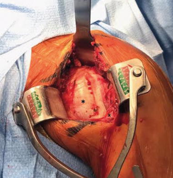
The skin incision is generally 8–10 cm in length and begins near the lateral margin of the coracoid process, extending distally along the deltopectoral groove toward the deltoid tuberosity. Following skin incision, the subcutaneous tissues are carefully dissected until the fascia overlying the deltoid and pectoralis major is identified.
The Cephalic Vein: Preserve or Mobilize?
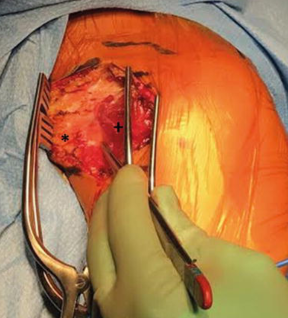
The cephalic vein is identified within the deltopectoral interval and carefully mobilized. It may be retracted either medially or laterally depending on the surgeon’s preference.
An anatomical study [1] of 40 cadaveric specimens demonstrated a greater number of feeder vessels entering the cephalic vein from the deltoid side than from the medial side. The mean number of lateral branches was 2.8 compared with 1.3 medial branches. These findings suggest that lateral retraction of the cephalic vein may reduce bleeding from its deltoid-side tributaries.
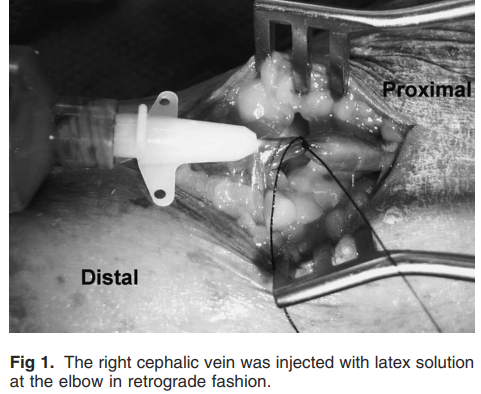
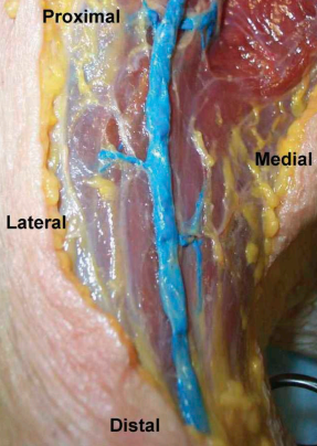
After mobilization of the vein, blunt dissection is continued between the deltoid and pectoralis major. A Kobel retractor can facilitate exposure of the clavipectoral fascia and conjoint tendon.
Protecting the Musculocutaneous and Axillary Nerves
Once the clavipectoral fascia is exposed, it is divided in line with the incision. The conjoint tendon is retracted medially to expose the subscapularis.
This is a critical stage of the approach. Excessive medial retraction of the conjoint tendon should be avoided because of the risk of musculocutaneous nerve neuropraxia. The axillary nerve must also be identified and protected, particularly because it courses close to the inferior border of the subscapularis.
The relationship of these nerves to the operative field becomes especially important during inferior capsular release and subscapularis mobilization.
The subscapularis lies directly anterior to the glenohumeral capsule and must be released to gain access to the joint.
Humeral Exposure
Once the capsule is isolated, a retractor is placed along the inferior glenoid to protect the axillary nerve. Capsulotomy is then continued along the anatomic neck and inferiorly down the humerus. Complete release of the capsule from the humeral neck until the latissimus dorsi tendon is visualized is an important step in improving glenoid exposure.
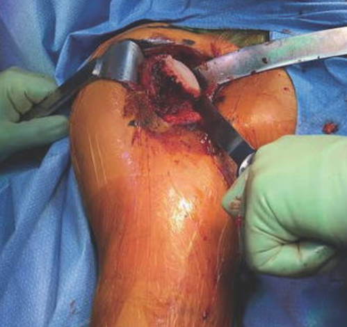
The humeral head can then be delivered by placing the arm in adduction, extension, and external rotation. Appropriate retractors protect the surrounding soft tissues during the humeral head cut. The level and angle of the cut should correspond to the intended implant geometry. The resected humeral head can also serve as a useful source of bone graft when required.
Glenoid Exposure
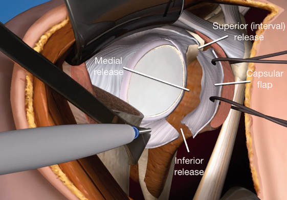
Glenoid exposure is facilitated by posterior, anterior, and inferior retractors. The Fukuda retractor retracts the humeral shaft posteriorly and inferiorly, while an anterior double-pronged retractor and inferior Hohmann retractor improve visualization and protect the axillary nerve.
The capsule, labrum, and remaining subscapularis tissue are released circumferentially to expose the glenoid. Inferior capsular release is particularly important. The rotator interval is released toward the base of the coracoid to improve exposure.
Care must be taken not to extend the anterior capsular release beyond the 6 o’clock position because of concerns regarding postoperative posterior instability.
Implant Preparation
Once the humerus and glenoid have been adequately exposed, glenoid preparation can begin. The guide pin is positioned approximately 11–12 mm above the inferior glenoid rim to facilitate appropriate component placement and minimize scapular notching. Concentric reaming is then performed over the guide pin.
For reverse shoulder arthroplasty, the central peg is prepared and the baseplate is secured with peripheral screws. On the humeral side, sequential canal reaming and broaching are performed until the appropriate implant size is achieved. Trial components are used to assess range of motion and stability before final implantation.
The final shoulder should be taken through a complete range of motion to ensure that the construct is stable without being overstuffed.
Closure
After the components have been appropriately placed and trialed, wound is irrigated with normal saline and subscapularis tendon is repaired
Advantages of the Deltopectoral Approach
The deltopectoral approach offers several important advantages:
1. Muscle-preserving exposure
The approach follows an internervous and intermuscular plane, preserving the origins of both the deltoid and pectoralis major and avoiding muscle splitting. This can reduce bleeding and minimize the risk of denervation.
2. Extensibility
If a fracture occurs distal to the humeral stem, the approach can readily be extended into an anterolateral approach to the humerus.
3. Excellent anterior and inferior access
The anterior trajectory provides particularly good access to inferior humeral osteophytes and the inferior capsule.
4. Favorable glenoid exposure
The inferior portion of the glenoid is readily accessible, facilitating glenoid component positioning.
Limitations and Potential Pitfalls
The principal limitation is the need to release and subsequently repair the subscapularis. In shoulder arthroplasty, subscapularis deficiency has been associated with increased instability, and failure of the subscapularis repair remains an important concern.
The anterior trajectory can also make access to posterior glenoid structures, the posterior capsule, and the greater tuberosity more difficult. This limitation becomes particularly relevant in proximal humeral fractures with large greater tuberosity fragments.
Practical Surgical Pearls
Several principles emerge from the surgical technique:
- Position the shoulder to allow extension before starting the approach.
- Identify and protect the cephalic vein and understand its tributary anatomy.
- Avoid aggressive medial retraction of the conjoint tendon to reduce the risk of musculocutaneous neuropraxia.
- Identify the axillary nerve before extensive inferior capsular release.
- Protect the anterior circumflex humeral vessels during inferior subscapularis release.
- Choose the subscapularis release technique deliberately, considering healing mechanism, tendon excursion, and postoperative external rotation.
- Avoid overtightening the subscapularis repair, as this can restrict external rotation.
- Obtain complete inferior and posterior glenoid exposure without excessive capsular release.
- Confirm stability and range of motion before finalizing the construct.
- Treat subscapularis repair as an integral component of the arthroplasty rather than simply the final closure step.
Take-Home Message
The deltopectoral approach provides reproducible and extensile access to the shoulder while preserving the major surrounding muscles. Its success depends less on the skin incision itself and more on understanding the three-dimensional anatomy of the cephalic vein, conjoint tendon, musculocutaneous nerve, axillary nerve, subscapularis, capsule, and glenoid.
Among the most important technical decisions is the method of subscapularis management.
Ultimately, meticulous exposure, protection of neurovascular structures, appropriate implant positioning, and a secure—but not overly tight—subscapularis repair are central to achieving a stable and functional shoulder arthroplasty through the deltopectoral approach.
For informative videos related to Shoulder problems and their treatment options, Sports Injuries and other orthopedic conditions, visit our YouTube channel Bangalore Shoulder Institute – https://www.youtube.com/@BangaloreShoulderInstitute
REFERENCES
- Radkowski CA, Richards RS, Pietrobon R, Moorman CT 3rd. An anatomic study of the cephalic vein in the deltopectoral shoulder approach. Clin Orthop Relat Res. 2006 Jan;442:139-42. doi: 10.1097/01.blo.0000181146.78434.da. PMID: 16394752.